1MYR

MYROSINASE FROM SINAPIS ALBA


Experimental Data Snapshot

  • Method: X-RAY DIFFRACTION
  • Resolution: 1.64 Å
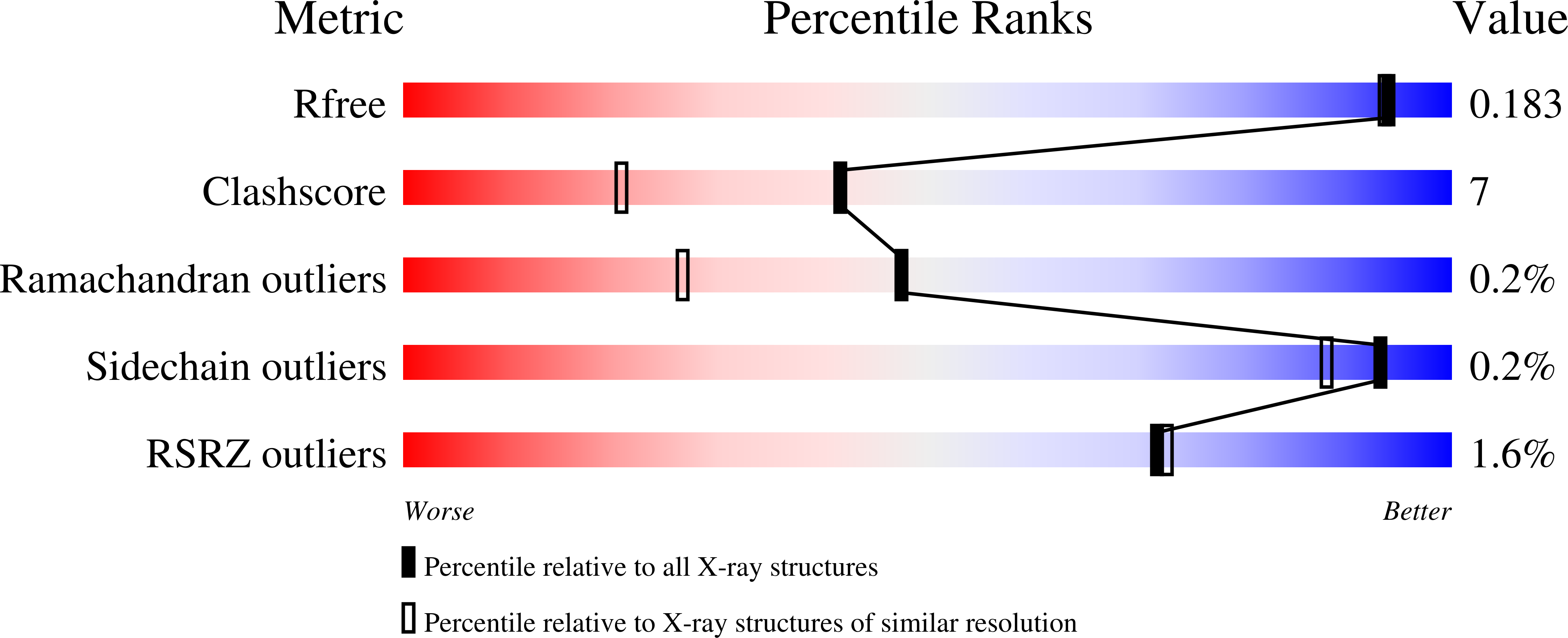
  • R-Value Free: 0.187 
  • R-Value Work: 0.152 
  • R-Value Observed: 0.152 

Starting Model: experimental
View more details

wwPDB Validation   3D Report Full Report


Ligand Structure Quality Assessment 


This is version 2.2 of the entry. See complete history


Literature

The crystal structures of Sinapis alba myrosinase and a covalent glycosyl-enzyme intermediate provide insights into the substrate recognition and active-site machinery of an S-glycosidase.

Burmeister, W.P.Cottaz, S.Driguez, H.Iori, R.Palmieri, S.Henrissat, B.

(1997) Structure 5: 663-675

  • DOI: https://doi.org/10.1016/s0969-2126(97)00221-9
  • Primary Citation of Related Structures:  
    1MYR

  • PubMed Abstract: 

    Myrosinase is the enzyme responsible for the hydrolysis of a variety of plant anionic 1-thio-beta-D-glucosides called glucosinolates. Myrosinase and glucosinolates, which are stored in different tissues of the plant, are mixed during mastication generating toxic by-products that are believed to play a role in the plant defence system. Whilst O-glycosidases are extremely widespread in nature, myrosinase is the only known S-glycosidase. This intriguing enzyme, which shows sequence similarities with O-glycosidases, offers the opportunity to analyze the similarities and differences between enzymes hydrolyzing S- and O-glycosidic bonds. The structures of native myrosinase from white mustard seed (Sinapis alba) and of a stable glycosyl-enzyme intermediate have been solved at 1.6 A resolution. The protein folds into a (beta/alpha)8-barrel structure, very similar to that of the cyanogenic beta-glucosidase from white clover. The enzyme forms a dimer stabilized by a Zn2+ ion and is heavily glycosylated. At one glycosylation site the complete structure of a plant-specific heptasaccharide is observed. The myrosinase structure reveals a hydrophobic pocket, ideally situated for the binding of the hydrophobic sidechain of glucosinolates, and two arginine residues positioned for interaction with the sulphate group of the substrate. With the exception of the replacement of the general acid/base glutamate by a glutamine residue, the catalytic machinery of myrosinase is identical to that of the cyanogenic beta-glucosidase. The structure of the glycosyl-enzyme intermediate shows that the sugar ring is bound via an alpha-glycosidic linkage to Glu409, the catalytic nucleophile of myrosinase. The structure of myrosinase shows features which illustrate the adaptation of the plant enzyme to the dehydrated environment of the seed. The catalytic mechanism of myrosinase is explained by the excellent leaving group properties of the substrate aglycons, which do not require the assistance of an enzymatic acid catalyst. The replacement of the general acid/base glutamate of O-glycosidases by a glutamine residue in myrosinase suggests that for hydrolysis of the glycosyl-enzyme, the role of this residue is to ensure a precise positioning of a water molecule rather than to provide general base assistance.


  • Organizational Affiliation

    European Synchrotron Radiation Facility (ESRF), BP 220, F-38043 Grenoble cedex, France. [email protected]


Macromolecules
Find similar proteins by:  (by identity cutoff)  |  3D Structure
Entity ID: 1
MoleculeChains Sequence LengthOrganismDetailsImage
MYROSINASE501Sinapis albaMutation(s): 0 
EC: 3.2.3.1 (PDB Primary Data), 3.2.1.147 (UniProt)
UniProt
Find proteins for P29736 (Sinapis alba)
Explore P29736 
Go to UniProtKB:  P29736
Entity Groups  
Sequence Clusters30% Identity50% Identity70% Identity90% Identity95% Identity100% Identity
UniProt GroupP29736
Glycosylation
Glycosylation Sites: 9
Sequence Annotations
Expand
  • Reference Sequence
Oligosaccharides

Help

Entity ID: 2
MoleculeChains Length2D Diagram Glycosylation3D Interactions
2-acetamido-2-deoxy-beta-D-glucopyranose-(1-4)-2-acetamido-2-deoxy-beta-D-glucopyranose
B, E
2N-Glycosylation
Glycosylation Resources
GlyTouCan:  G42666HT
GlyCosmos:  G42666HT
GlyGen:  G42666HT
Entity ID: 3
MoleculeChains Length2D Diagram Glycosylation3D Interactions
beta-D-xylopyranose-(1-2)-beta-D-mannopyranose-(1-4)-2-acetamido-2-deoxy-beta-D-glucopyranose-(1-4)-[alpha-L-fucopyranose-(1-3)]2-acetamido-2-deoxy-beta-D-glucopyranose
C
5N-Glycosylation
Glycosylation Resources
GlyTouCan:  G06206UV
GlyCosmos:  G06206UV
GlyGen:  G06206UV
Entity ID: 4
MoleculeChains Length2D Diagram Glycosylation3D Interactions
beta-D-xylopyranose-(1-2)-[alpha-D-mannopyranose-(1-3)][alpha-D-mannopyranose-(1-6)]beta-D-mannopyranose-(1-4)-2-acetamido-2-deoxy-beta-D-glucopyranose-(1-4)-[alpha-L-fucopyranose-(1-3)]2-acetamido-2-deoxy-beta-D-glucopyranose
D
7N-Glycosylation
Glycosylation Resources
GlyTouCan:  G26295XE
GlyCosmos:  G26295XE
GlyGen:  G26295XE
Small Molecules
Ligands 4 Unique
IDChains Name / Formula / InChI Key2D Diagram3D Interactions
NAG
Query on NAG

Download Ideal Coordinates CCD File 
F [auth A],
G [auth A],
H [auth A],
I [auth A],
J [auth A]
2-acetamido-2-deoxy-beta-D-glucopyranose
C8 H15 N O6
OVRNDRQMDRJTHS-FMDGEEDCSA-N
SO4
Query on SO4

Download Ideal Coordinates CCD File 
L [auth A]
M [auth A]
N [auth A]
O [auth A]
P [auth A]
L [auth A],
M [auth A],
N [auth A],
O [auth A],
P [auth A],
Q [auth A],
R [auth A],
S [auth A],
T [auth A],
U [auth A]
SULFATE ION
O4 S
QAOWNCQODCNURD-UHFFFAOYSA-L
GOL
Query on GOL

Download Ideal Coordinates CCD File 
V [auth A],
W [auth A],
X [auth A],
Y [auth A]
GLYCEROL
C3 H8 O3
PEDCQBHIVMGVHV-UHFFFAOYSA-N
ZN
Query on ZN

Download Ideal Coordinates CCD File 
K [auth A]ZINC ION
Zn
PTFCDOFLOPIGGS-UHFFFAOYSA-N
Experimental Data & Validation

Experimental Data

  • Method: X-RAY DIFFRACTION
  • Resolution: 1.64 Å
  • R-Value Free: 0.187 
  • R-Value Work: 0.152 
  • R-Value Observed: 0.152 
  • Space Group: C 2 2 21
Unit Cell:
Length ( Å )Angle ( ˚ )
a = 134.3α = 90
b = 136.4β = 90
c = 80.3γ = 90
Software Package:
Software NamePurpose
MOSFLMdata reduction
ROTAVATAdata reduction
Agrovatadata reduction
X-PLORmodel building
X-PLORrefinement
CCP4data scaling
ROTAVATAdata scaling
X-PLORphasing

Structure Validation

View Full Validation Report


Ligand Structure Quality Assessment 


Entry History 

Deposition Data

Revision History  (Full details and data files)

  • Version 1.0: 1997-06-16
    Type: Initial release
  • Version 1.1: 2008-03-24
    Changes: Version format compliance
  • Version 1.2: 2011-07-13
    Changes: Derived calculations, Version format compliance
  • Version 1.3: 2011-11-16
    Changes: Atomic model
  • Version 2.0: 2020-07-29
    Type: Remediation
    Reason: Carbohydrate remediation
    Changes: Advisory, Atomic model, Data collection, Derived calculations, Non-polymer description, Refinement description, Structure summary
  • Version 2.1: 2023-08-09
    Changes: Database references, Derived calculations, Refinement description, Structure summary
  • Version 2.2: 2024-10-23
    Changes: Data collection, Structure summary