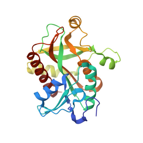
1ODJ

PURINE NUCLEOSIDE PHOSPHORYLASE FROM THERMUS THERMOPHILUS


Experimental Data Snapshot

  • Method: X-RAY DIFFRACTION
  • Resolution: 2.40 Å
  • R-Value Free: 0.286 
  • R-Value Work: 0.221 
  • R-Value Observed: 0.221 

Starting Model: experimental
View more details

wwPDB Validation   3D Report Full Report


Ligand Structure Quality Assessment 


This is version 1.4 of the entry. See complete history


Literature

Crystal Structure of Purine Nucleoside Phosphorylase from Thermus Thermophilus

Tahirov, T.H.Inagaki, E.Ohshima, N.Kitao, T.Kuroishi, C.Ukita, Y.Takio, K.Kobayashi, M.Kuramitsu, S.Yokoyama, S.Miyano, M.

(2004) J Mol Biol 337: 1149

  • DOI: https://doi.org/10.1016/j.jmb.2004.02.016
  • Primary Citation of Related Structures:  
    1ODI, 1ODJ, 1ODK, 1ODL

  • PubMed Abstract: 

    The purine nucleoside phosphorylase from Thermus thermophilus crystallized in space group P4(3)2(1)2 with the unit cell dimensions a = 131.9 A and c = 169.9 A and one biologically active hexamer in the asymmetric unit. The structure was solved by the molecular replacement method and refined at a 1.9A resolution to an r(free) value of 20.8%. The crystals of the binary complex with sulfate ion and ternary complexes with sulfate and adenosine or guanosine were also prepared and their crystal structures were refined at 2.1A, 2.4A and 2.4A, respectively. The overall structure of the T.thermophilus enzyme is similar to the structures of hexameric enzymes from Escherichia coli and Sulfolobus solfataricus, but significant differences are observed in the purine base recognition site. A base recognizing aspartic acid, which is conserved among the hexameric purine nucleoside phosphorylases, is Asn204 in the T.thermophilus enzyme, which is reminiscent of the base recognizing asparagine in trimeric purine nucleoside phosphorylases. Isothermal titration calorimetry measurements indicate that both adenosine and guanosine bind the enzyme with nearly similar affinity. However, the functional assays show that as in trimeric PNPs, only the guanosine is a true substrate of the T.thermophilus enzyme. In the case of adenosine recognition, the Asn204 forms hydrogen bonds with N6 and N7 of the base. While in the case of guanosine recognition, the Asn204 is slightly shifted together with the beta(9)alpha(7) loop and predisposed to hydrogen bond formation with O6 of the base in the transition state. The obtained experimental data suggest that the catalytic properties of the T.thermophilus enzyme are reminiscent of the trimeric rather than hexameric purine nucleoside phosphorylases.


  • Organizational Affiliation

    Highthroughput Factory, RIKEN Harima Institute, 1-1-1 Kouto, Mikazuki-cho, Sayo-gun, Hyogo 679-5148, Japan. [email protected]


Macromolecules
Find similar proteins by:  (by identity cutoff)  |  3D Structure
Entity ID: 1
MoleculeChains Sequence LengthOrganismDetailsImage
PURINE NUCLEOSIDE PHOSPHORYLASE
A, B, C, D, E
A, B, C, D, E, F
235Thermus thermophilus HB8Mutation(s): 0 
EC: 2.4.2.28 (PDB Primary Data), 2.4.2.3 (UniProt)
UniProt
Find proteins for Q5SID9 (Thermus thermophilus (strain ATCC 27634 / DSM 579 / HB8))
Explore Q5SID9 
Go to UniProtKB:  Q5SID9
Entity Groups  
Sequence Clusters30% Identity50% Identity70% Identity90% Identity95% Identity100% Identity
UniProt GroupQ5SID9
Sequence Annotations
Expand
  • Reference Sequence
Small Molecules
Ligands 2 Unique
IDChains Name / Formula / InChI Key2D Diagram3D Interactions
GMP
Query on GMP

Download Ideal Coordinates CCD File 
H [auth A]
J [auth B]
L [auth C]
N [auth D]
P [auth E]
H [auth A],
J [auth B],
L [auth C],
N [auth D],
P [auth E],
R [auth F]
GUANOSINE
C10 H13 N5 O5
NYHBQMYGNKIUIF-UUOKFMHZSA-N
SO4
Query on SO4

Download Ideal Coordinates CCD File 
G [auth A]
I [auth B]
K [auth C]
M [auth D]
O [auth E]
G [auth A],
I [auth B],
K [auth C],
M [auth D],
O [auth E],
Q [auth F]
SULFATE ION
O4 S
QAOWNCQODCNURD-UHFFFAOYSA-L
Binding Affinity Annotations 
IDSourceBinding Affinity
GMP PDBBind:  1ODJ Kd: 8.00e+4 (nM) from 1 assay(s)
Experimental Data & Validation

Experimental Data

  • Method: X-RAY DIFFRACTION
  • Resolution: 2.40 Å
  • R-Value Free: 0.286 
  • R-Value Work: 0.221 
  • R-Value Observed: 0.221 
  • Space Group: P 43 21 2
Unit Cell:
Length ( Å )Angle ( ˚ )
a = 132.757α = 90
b = 132.757β = 90
c = 172.06γ = 90
Software Package:
Software NamePurpose
CNSrefinement
HKL-2000data reduction
HKL-2000data scaling
CNSphasing

Structure Validation

View Full Validation Report


Ligand Structure Quality Assessment 


Entry History 

Deposition Data

Revision History  (Full details and data files)

  • Version 1.0: 2003-03-04
    Type: Initial release
  • Version 1.1: 2011-05-08
    Changes: Version format compliance
  • Version 1.2: 2011-07-13
    Changes: Version format compliance
  • Version 1.3: 2019-05-08
    Changes: Data collection, Experimental preparation, Other
  • Version 1.4: 2024-05-01
    Changes: Data collection, Database references, Other, Refinement description