1ODV

Photoactive yellow protein 1-25 deletion mutant


Experimental Data Snapshot

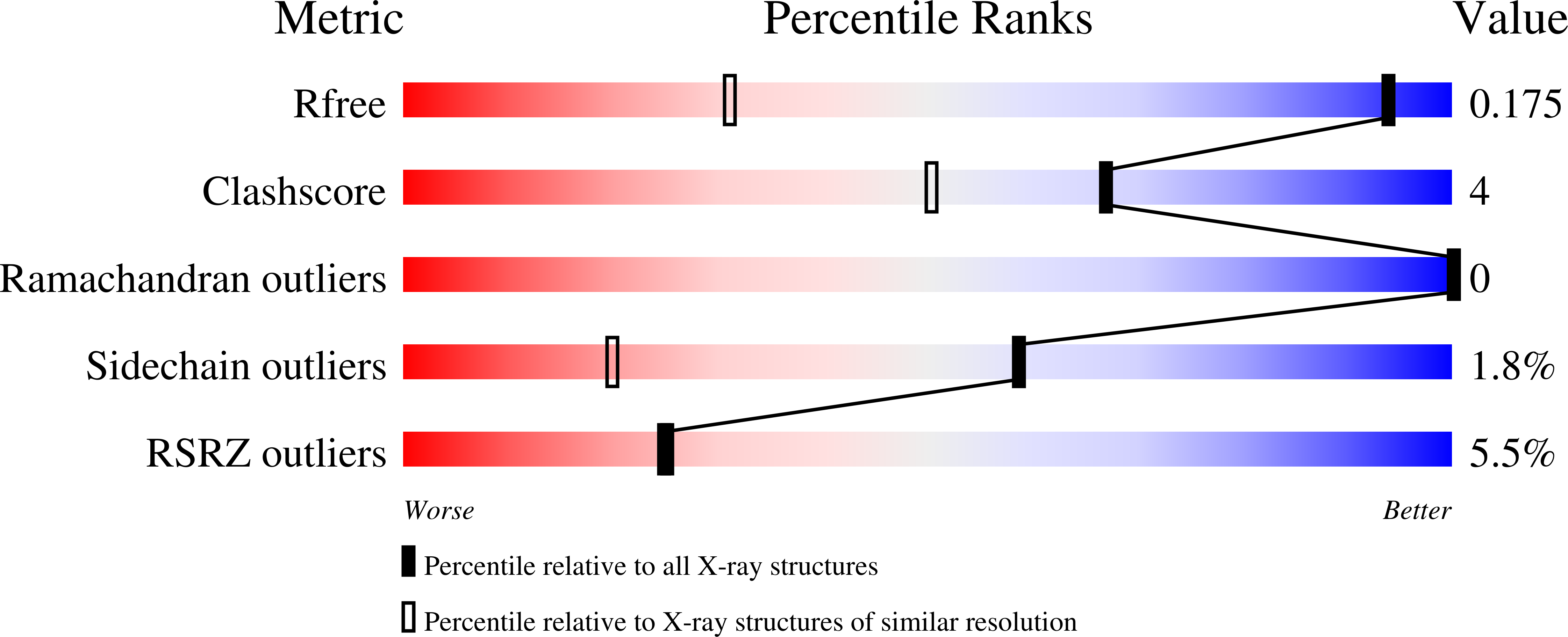
  • Method: X-RAY DIFFRACTION
  • Resolution: 1.14 Å
  • R-Value Free: 0.177 
  • R-Value Observed: 0.147 

Starting Model: experimental
View more details

wwPDB Validation   3D Report Full Report


Ligand Structure Quality Assessment 


This is version 1.4 of the entry. See complete history


Literature

Pas Domains.Common Structure and Common Flexibility

Vreede, J.Van Der Horst, M.A.Hellingwerf, K.J.Crielaard, W.Van Aalten, D.M.F.

(2003) J Biol Chem 278: 18434

  • DOI: https://doi.org/10.1074/jbc.M301701200
  • Primary Citation of Related Structures:  
    1ODV

  • PubMed Abstract: 

    PAS (PER-ARNT-SIM) domains are a family of sensor protein domains involved in signal transduction in a wide range of organisms. Recent structural studies have revealed that these domains contain a structurally conserved alpha/beta-fold, whereas almost no conservation is observed at the amino acid sequence level. The photoactive yellow protein, a bacterial light sensor, has been proposed as the PAS structural prototype yet contains an N-terminal helix-turn-helix motif not found in other PAS domains. Here we describe the atomic resolution structure of a photoactive yellow protein deletion mutant lacking this motif, revealing that the PAS domain is indeed able to fold independently and is not affected by the removal of these residues. Computer simulations of currently known PAS domain structures reveal that these domains are not only structurally conserved but are also similar in their conformational flexibilities. The observed motions point to a possible common mechanism for communicating ligand binding/activation to downstream transducer proteins.


  • Organizational Affiliation

    Department of Microbiology, Swammerdam Institute for Life Sciences, University of Amsterdam, 1018WV, Amsterdam, The Netherlands.


Macromolecules
Find similar proteins by:  (by identity cutoff)  |  3D Structure
Entity ID: 1
MoleculeChains Sequence LengthOrganismDetailsImage
PHOTOACTIVE YELLOW PROTEIN
A, B
100Halorhodospira halophilaMutation(s): 0 
UniProt
Find proteins for P16113 (Halorhodospira halophila)
Explore P16113 
Go to UniProtKB:  P16113
Entity Groups  
Sequence Clusters30% Identity50% Identity70% Identity90% Identity95% Identity100% Identity
UniProt GroupP16113
Sequence Annotations
Expand
  • Reference Sequence
Experimental Data & Validation

Experimental Data

  • Method: X-RAY DIFFRACTION
  • Resolution: 1.14 Å
  • R-Value Free: 0.177 
  • R-Value Observed: 0.147 
  • Space Group: P 43 21 2
Unit Cell:
Length ( Å )Angle ( ˚ )
a = 82.566α = 90
b = 82.566β = 90
c = 63.453γ = 90
Software Package:
Software NamePurpose
SHELXL-97refinement
DENZOdata reduction
SCALEPACKdata scaling
SHELXphasing

Structure Validation

View Full Validation Report


Ligand Structure Quality Assessment 


Entry History 

Deposition Data

Revision History  (Full details and data files)

  • Version 1.0: 2003-03-18
    Type: Initial release
  • Version 1.1: 2011-05-08
    Changes: Version format compliance
  • Version 1.2: 2011-07-13
    Changes: Version format compliance
  • Version 1.3: 2019-05-22
    Changes: Advisory, Data collection, Derived calculations, Other, Refinement description
  • Version 1.4: 2023-12-13
    Changes: Advisory, Data collection, Database references, Derived calculations, Other, Refinement description