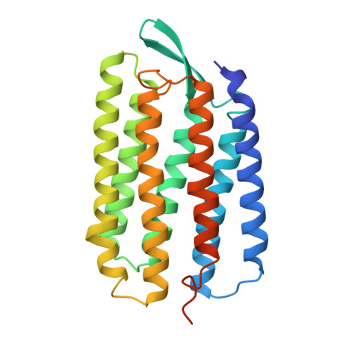
1QHJ

X-RAY STRUCTURE OF BACTERIORHODOPSIN GROWN IN LIPIDIC CUBIC PHASES


Experimental Data Snapshot

  • Method: X-RAY DIFFRACTION
  • Resolution: 1.90 Å
  • R-Value Free: 0.245 
  • R-Value Work: 0.224 

Starting Model: experimental
View more details

wwPDB Validation   3D Report Full Report


Ligand Structure Quality Assessment 


This is version 1.4 of the entry. See complete history


Literature

Protein, lipid and water organization in bacteriorhodopsin crystals: a molecular view of the purple membrane at 1.9 A resolution.

Belrhali, H.Nollert, P.Royant, A.Menzel, C.Rosenbusch, J.P.Landau, E.M.Pebay-Peyroula, E.

(1999) Structure 7: 909-917

  • DOI: https://doi.org/10.1016/s0969-2126(99)80118-x
  • Primary Citation of Related Structures:  
    1QHJ

  • PubMed Abstract: 

    Bacteriorhodopsin (bR) from Halobacterium salinarum is a proton pump that converts the energy of light into a proton gradient that drives ATP synthesis. The protein comprises seven transmembrane helices and in vivo is organized into purple patches, in which bR and lipids form a crystalline two-dimensional array. Upon absorption of a photon, retinal, which is covalently bound to Lys216 via a Schiff base, is isomerized to a 13-cis,15-anti configuration. This initiates a sequence of events - the photocycle - during which a proton is transferred from the Schiff base to Asp85, followed by proton release into the extracellular medium and reprotonation from the cytoplasmic side. The structure of bR in the ground state was solved to 1.9 A resolution from non-twinned crystals grown in a lipidic cubic phase. The structure reveals eight well-ordered water molecules in the extracellular half of the putative proton translocation pathway. The water molecules form a continuous hydrogen-bond network from the Schiff-base nitrogen (Lys216) to Glu194 and Glu204 and includes residues Asp85, Asp212 and Arg82. This network is involved both in proton translocation occurring during the photocycle, as well as in stabilizing the structure of the ground state. Nine lipid phytanyl moieties could be modeled into the electron-density maps. Matrix-assisted laser desorption/ionization mass spectrometry (MALDI-MS) analysis of single crystals demonstrated the presence of four different charged lipid species. The structure of protein, lipid and water molecules in the crystals represents the functional entity of bR in the purple membrane of the bacteria at atomic resolution. Proton translocation from the Schiff base to the extracellular medium is mediated by a hydrogen-bond network that involves charged residues and water molecules.


  • Organizational Affiliation

    ESRF, Grenoble, France.


Macromolecules
Find similar proteins by:  (by identity cutoff)  |  3D Structure
Entity ID: 1
MoleculeChains Sequence LengthOrganismDetailsImage
PROTEIN (BACTERIORHODOPSIN)248Halobacterium salinarumMutation(s): 0 
Membrane Entity: Yes 
UniProt
Find proteins for P02945 (Halobacterium salinarum (strain ATCC 700922 / JCM 11081 / NRC-1))
Explore P02945 
Go to UniProtKB:  P02945
Entity Groups  
Sequence Clusters30% Identity50% Identity70% Identity90% Identity95% Identity100% Identity
UniProt GroupP02945
Sequence Annotations
Expand
  • Reference Sequence
Small Molecules
Ligands 2 Unique
IDChains Name / Formula / InChI Key2D Diagram3D Interactions
PH1
Query on PH1

Download Ideal Coordinates CCD File 
C [auth A]
D [auth A]
E [auth A]
F [auth A]
G [auth A]
C [auth A],
D [auth A],
E [auth A],
F [auth A],
G [auth A],
H [auth A],
I [auth A],
J [auth A],
K [auth A]
1,2-[DI-2,6,10,14-TETRAMETHYL-HEXADECAN-16-OXY]-PROPANE
C43 H88 O2
ICKFQWWHLUUVMF-FYYCEQFTSA-N
RET
Query on RET

Download Ideal Coordinates CCD File 
B [auth A]RETINAL
C20 H28 O
NCYCYZXNIZJOKI-OVSJKPMPSA-N
Experimental Data & Validation

Experimental Data

  • Method: X-RAY DIFFRACTION
  • Resolution: 1.90 Å
  • R-Value Free: 0.245 
  • R-Value Work: 0.224 
  • Space Group: P 63
Unit Cell:
Length ( Å )Angle ( ˚ )
a = 60.8α = 90
b = 60.8β = 90
c = 110.52γ = 120
Software Package:
Software NamePurpose
BASEDmodel building
CNSrefinement
XDSdata reduction
CCP4data scaling
BASEDphasing

Structure Validation

View Full Validation Report


Ligand Structure Quality Assessment 


Entry History 

Deposition Data

Revision History  (Full details and data files)

  • Version 1.0: 1999-07-21
    Type: Initial release
  • Version 1.1: 2007-10-16
    Changes: Version format compliance
  • Version 1.2: 2011-07-13
    Changes: Derived calculations, Version format compliance
  • Version 1.3: 2023-08-16
    Changes: Data collection, Database references, Derived calculations, Refinement description
  • Version 1.4: 2024-10-30
    Changes: Structure summary