1R37

Alcohol dehydrogenase from sulfolobus solfataricus complexed with NAD(H) and 2-ethoxyethanol


Experimental Data Snapshot

  • Method: X-RAY DIFFRACTION
  • Resolution: 2.30 Å
  • R-Value Free: 0.225 
  • R-Value Work: 0.193 

Starting Model: experimental
View more details

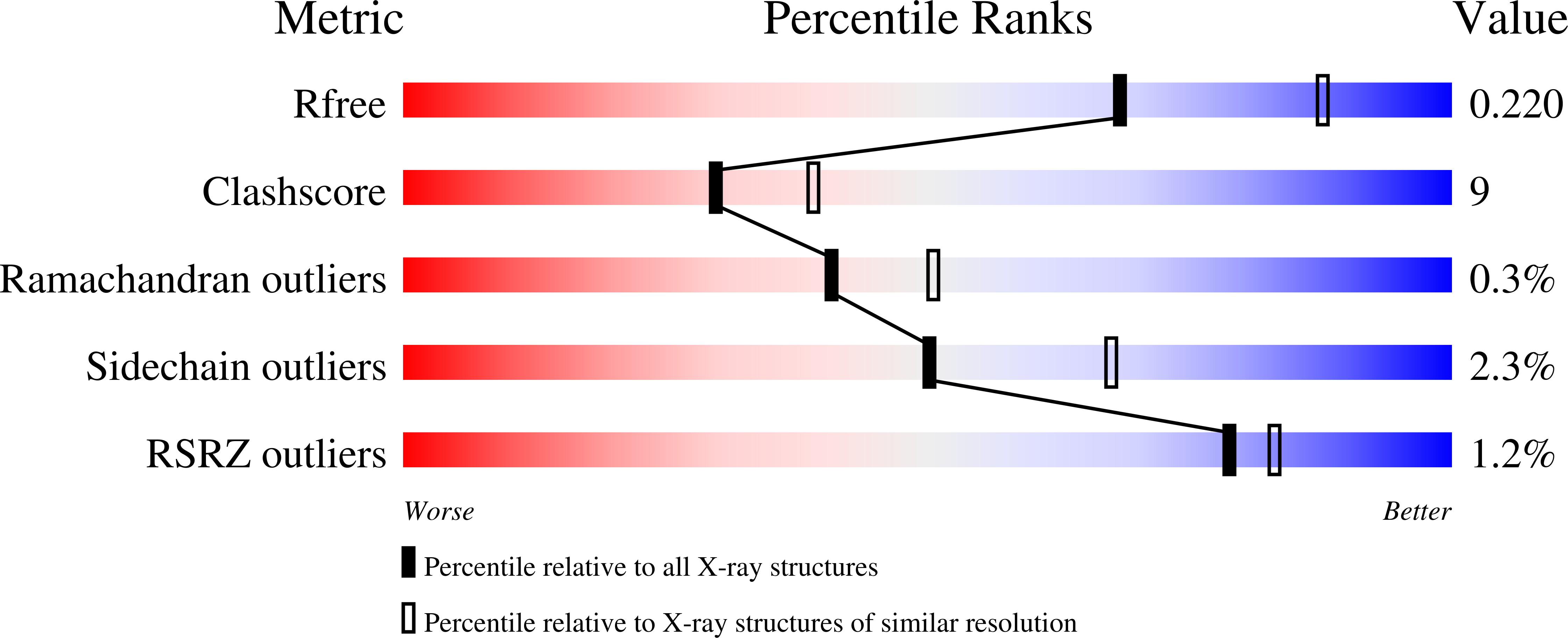
wwPDB Validation   3D Report Full Report


Ligand Structure Quality Assessment 


This is version 1.3 of the entry. See complete history


Literature

Crystal structure of a ternary complex of the alcohol dehydrogenase from Sulfolobus solfataricus

Esposito, L.Bruno, I.Sica, F.Raia, C.A.Giordano, A.Rossi, M.Mazzarella, L.Zagari, A.

(2003) Biochemistry 42: 14397-14407

  • DOI: https://doi.org/10.1021/bi035271b
  • Primary Citation of Related Structures:  
    1R37

  • PubMed Abstract: 

    The crystal structure of a ternary complex of the alcohol dehydrogenase from the archaeon Sulfolobus solfataricus (SsADH) has been determined at 2.3 A. The asymmetric unit contains a dimer with a NADH and a 2-ethoxyethanol molecule bound to each subunit. The comparison with the apo structure of the enzyme reveals that this medium chain ADH undergoes a substantial conformational change in the apo-holo transition, accompanied by loop movements at the domain interface. The extent of domain closure is similar to that observed for the classical horse liver ADH, although some differences are found which can be related to the different oligomeric states of the enzymes. Compared to its apo form, the SsADH ternary complex shows a change in the ligation state of the active site zinc ion which is no longer bound to Glu69, providing additional evidence of the dynamic role played by the conserved glutamate residue in ADHs. In addition, the structure presented here allows the identification of the substrate site and hence of the residues that are important in the binding of both the substrate and the coenzyme.


  • Organizational Affiliation

    Istituto di Biostrutture e Bioimmagini, CNR, via Mezzocannone 6-8, I-80134 Napoli, Italy.


Macromolecules
Find similar proteins by:  (by identity cutoff)  |  3D Structure
Entity ID: 1
MoleculeChains Sequence LengthOrganismDetailsImage
NAD-dependent alcohol dehydrogenase
A, B
347Saccharolobus solfataricusMutation(s): 0 
Gene Names: ADH or SSO2536
EC: 1.1.1.1
UniProt
Find proteins for P39462 (Saccharolobus solfataricus (strain ATCC 35092 / DSM 1617 / JCM 11322 / P2))
Explore P39462 
Go to UniProtKB:  P39462
Entity Groups  
Sequence Clusters30% Identity50% Identity70% Identity90% Identity95% Identity100% Identity
UniProt GroupP39462
Sequence Annotations
Expand
  • Reference Sequence
Small Molecules
Ligands 3 Unique
IDChains Name / Formula / InChI Key2D Diagram3D Interactions
NAD
Query on NAD

Download Ideal Coordinates CCD File 
E [auth A],
I [auth B]
NICOTINAMIDE-ADENINE-DINUCLEOTIDE
C21 H27 N7 O14 P2
BAWFJGJZGIEFAR-NNYOXOHSSA-N
ETX
Query on ETX

Download Ideal Coordinates CCD File 
F [auth A],
J [auth B]
2-ETHOXYETHANOL
C4 H10 O2
ZNQVEEAIQZEUHB-UHFFFAOYSA-N
ZN
Query on ZN

Download Ideal Coordinates CCD File 
C [auth A],
D [auth A],
G [auth B],
H [auth B]
ZINC ION
Zn
PTFCDOFLOPIGGS-UHFFFAOYSA-N
Experimental Data & Validation

Experimental Data

  • Method: X-RAY DIFFRACTION
  • Resolution: 2.30 Å
  • R-Value Free: 0.225 
  • R-Value Work: 0.193 
  • Space Group: C 1 2 1
Unit Cell:
Length ( Å )Angle ( ˚ )
a = 131.064α = 90
b = 83.014β = 96.46
c = 68.674γ = 90
Software Package:
Software NamePurpose
DENZOdata reduction
SCALEPACKdata scaling
AMoREphasing
CNSrefinement

Structure Validation

View Full Validation Report


Ligand Structure Quality Assessment 


Entry History 

Deposition Data

Revision History  (Full details and data files)

  • Version 1.0: 2004-02-10
    Type: Initial release
  • Version 1.1: 2007-10-16
    Changes: Version format compliance
  • Version 1.2: 2011-07-13
    Changes: Version format compliance
  • Version 1.3: 2023-10-25
    Changes: Data collection, Database references, Derived calculations, Refinement description