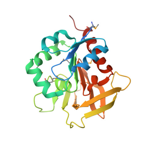
1R9G

Three-dimensional Structure of YaaE from Bacillus subtilis


Experimental Data Snapshot

  • Method: X-RAY DIFFRACTION
  • Resolution: 2.50 Å
  • R-Value Free: 0.279 
  • R-Value Work: 0.240 
  • R-Value Observed: 0.244 

wwPDB Validation   3D Report Full Report


This is version 1.3 of the entry. See complete history


Literature

Three-dimensional Structure of YaaE from Bacillus subtilis, a Glutaminase Implicated in Pyridoxal-5'-phosphate Biosynthesis.

Bauer, J.A.Bennett, E.M.Begley, T.P.Ealick, S.E.

(2004) J Biol Chem 279: 2704-2711

  • DOI: https://doi.org/10.1074/jbc.M310311200
  • Primary Citation of Related Structures:  
    1R9G

  • PubMed Abstract: 

    The structure of YaaE from Bacillus subtilis was determined at 2.5-A resolution. YaaE is a member of the triad glutamine aminotransferase family and functions in a recently identified alternate pathway for the biosynthesis of vitamin B(6). Proposed active residues include conserved Cys-79, His-170, and Glu-172. YaaE shows similarity to HisH, a glutaminase involved in histidine biosynthesis. YaaD associates with YaaE. A homology model of this protein was constructed. YaaD is predicted to be a (beta/alpha)(8) barrel on the basis of sequence comparisons. The predicted active site includes highly conserved residues 211-216 and 233-235. Finally, a homology model of a putative YaaD-YaaE complex was prepared using the structure of HisH-F as a model. This model predicts that the ammonia molecule generated by YaaE is channeled through the center of the YaaD barrel to the putative YaaD active site.


  • Organizational Affiliation

    Department of Chemistry and Chemical Biology, Cornell University, Ithaca, New York 14853, USA.


Macromolecules
Find similar proteins by:  (by identity cutoff)  |  3D Structure
Entity ID: 1
MoleculeChains Sequence LengthOrganismDetailsImage
Hypothetical protein yaaE
A, B
216Bacillus subtilisMutation(s): 5 
Gene Names: YAAEBSU00120
EC: 4.3.3.6 (UniProt), 3.5.1.2 (UniProt)
UniProt
Find proteins for P37528 (Bacillus subtilis (strain 168))
Explore P37528 
Go to UniProtKB:  P37528
Entity Groups  
Sequence Clusters30% Identity50% Identity70% Identity90% Identity95% Identity100% Identity
UniProt GroupP37528
Sequence Annotations
Expand
  • Reference Sequence
Small Molecules
Modified Residues  1 Unique
IDChains TypeFormula2D DiagramParent
MSE
Query on MSE
A, B
L-PEPTIDE LINKINGC5 H11 N O2 SeMET
Experimental Data & Validation

Experimental Data

  • Method: X-RAY DIFFRACTION
  • Resolution: 2.50 Å
  • R-Value Free: 0.279 
  • R-Value Work: 0.240 
  • R-Value Observed: 0.244 
  • Space Group: P 21 21 21
Unit Cell:
Length ( Å )Angle ( ˚ )
a = 45.5α = 90
b = 80.87β = 90
c = 115.39γ = 90
Software Package:
Software NamePurpose
MOSFLMdata reduction
SCALAdata scaling
CNSrefinement
CCP4data scaling
CNSphasing

Structure Validation

View Full Validation Report


Entry History 

Deposition Data

Revision History  (Full details and data files)

  • Version 1.0: 2004-01-27
    Type: Initial release
  • Version 1.1: 2008-04-29
    Changes: Version format compliance
  • Version 1.2: 2011-07-13
    Changes: Version format compliance
  • Version 1.3: 2024-10-30
    Changes: Data collection, Database references, Derived calculations, Structure summary