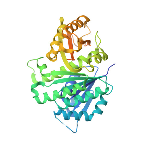
1RQ7

MYCOBACTERIUM TUBERCULOSIS FTSZ IN COMPLEX WITH GDP


Experimental Data Snapshot

  • Method: X-RAY DIFFRACTION
  • Resolution: 2.60 Å
  • R-Value Free: 0.242 
  • R-Value Work: 0.184 
  • R-Value Observed: 0.187 

Starting Model: experimental
View more details

wwPDB Validation   3D Report Full Report


Ligand Structure Quality Assessment 


This is version 1.5 of the entry. See complete history


Literature

Structure of Mycobacterium tuberculosis FtsZ reveals unexpected, G protein-like conformational switches.

Leung, A.K.Lucile White, E.Ross, L.J.Reynolds, R.C.DeVito, J.A.Borhani, D.W.

(2004) J Mol Biol 342: 953-970

  • DOI: https://doi.org/10.1016/j.jmb.2004.07.061
  • Primary Citation of Related Structures:  
    1RLU, 1RQ2, 1RQ7

  • PubMed Abstract: 

    We report three crystal structures of the Mycobacterium tuberculosis cell division protein FtsZ, as the citrate, GDP, and GTPgammaS complexes, determined at 1.89, 2.60, and 2.08A resolution. MtbFtsZ crystallized as a tight, laterally oriented dimer distinct from the longitudinal polymer observed for alphabeta-tubulin. Mutational data on Escherichia coli FtsZ suggest that this dimer interface is important for proper protofilament and "Z-ring" assembly and function. An alpha-to-beta secondary structure conformational switch at the dimer interface is spatially analogous to, and has many of the hallmarks of, the Switch I conformational changes exhibited by G-proteins upon activation. The presence of a gamma-phosphate in the FtsZ active site modulates the conformation of the "tubulin" loop T3 (spatially analogous to the G-protein Switch II); T3 switching upon gamma-phosphate ligation is directly coupled to the alpha-to-beta switch by steric overlap. The dual conformational switches observed here for the first time in an FtsZ link GTP binding and hydrolysis to FtsZ (and tubulin) lateral assembly and Z-ring contraction, and they are suggestive of an underappreciated functional analogy between FtsZ, tubulin and G-proteins.


  • Organizational Affiliation

    Drug Discovery Division, Southern Research Institute, Birmingham, AL 35205, USA.


Macromolecules
Find similar proteins by:  (by identity cutoff)  |  3D Structure
Entity ID: 1
MoleculeChains Sequence LengthOrganismDetailsImage
Cell division protein ftsZ
A, B
382Mycobacterium tuberculosisMutation(s): 0 
Gene Names: FTSZRV2150CMT2209MTCY270.18MB2174C
UniProt
Find proteins for P9WN95 (Mycobacterium tuberculosis (strain ATCC 25618 / H37Rv))
Explore P9WN95 
Go to UniProtKB:  P9WN95
Entity Groups  
Sequence Clusters30% Identity50% Identity70% Identity90% Identity95% Identity100% Identity
UniProt GroupP9WN95
Sequence Annotations
Expand
  • Reference Sequence
Small Molecules
Ligands 1 Unique
IDChains Name / Formula / InChI Key2D Diagram3D Interactions
GDP
Query on GDP

Download Ideal Coordinates CCD File 
C [auth A]GUANOSINE-5'-DIPHOSPHATE
C10 H15 N5 O11 P2
QGWNDRXFNXRZMB-UUOKFMHZSA-N
Experimental Data & Validation

Experimental Data

  • Method: X-RAY DIFFRACTION
  • Resolution: 2.60 Å
  • R-Value Free: 0.242 
  • R-Value Work: 0.184 
  • R-Value Observed: 0.187 
  • Space Group: P 65
Unit Cell:
Length ( Å )Angle ( ˚ )
a = 88.619α = 90
b = 88.619β = 90
c = 179.196γ = 120
Software Package:
Software NamePurpose
REFMACrefinement
SCALAdata scaling
TRUNCATEdata reduction
X-PLORmodel building
X-PLORrefinement
CCP4data scaling
TRUNCATEdata scaling
X-PLORphasing
MOSFLMdata reduction

Structure Validation

View Full Validation Report


Ligand Structure Quality Assessment 


Entry History 

Deposition Data

Revision History  (Full details and data files)

  • Version 1.0: 2004-08-31
    Type: Initial release
  • Version 1.1: 2008-04-29
    Changes: Version format compliance
  • Version 1.2: 2011-07-13
    Changes: Version format compliance
  • Version 1.3: 2017-10-11
    Changes: Refinement description
  • Version 1.4: 2019-07-24
    Changes: Data collection, Refinement description
  • Version 1.5: 2023-08-23
    Changes: Data collection, Database references, Derived calculations, Refinement description