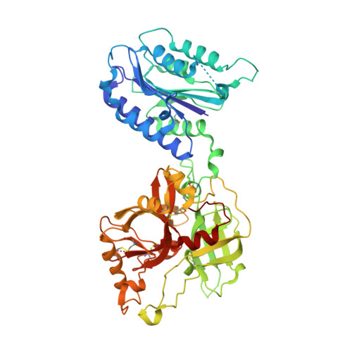
1RRK

Crystal Structure Analysis of the Bb segment of Factor B


Experimental Data Snapshot

  • Method: X-RAY DIFFRACTION
  • Resolution: 2.00 Å
  • R-Value Free: 0.245 
  • R-Value Work: 0.220 
  • R-Value Observed: 0.220 

Starting Model: experimental
View more details

wwPDB Validation   3D Report Full Report


This is version 1.5 of the entry. See complete history


Literature

Structural analysis of engineered Bb fragment of complement factor B: insights into the activation mechanism of the alternative pathway C3-convertase.

Ponnuraj, K.Xu, Y.Macon, K.Moore, D.Volanakis, J.E.Narayana, S.V.

(2004) Mol Cell 14: 17-28

  • DOI: https://doi.org/10.1016/s1097-2765(04)00160-1
  • Primary Citation of Related Structures:  
    1RRK, 1RS0, 1RTK

  • PubMed Abstract: 

    The C-terminal fragment, Bb, of factor B combines with C3b to form the pivotal C3-convertase, C3bBb, of alternative complement pathway. Bb consists of a von Willebrand factor type A (vWFA) domain that is structurally similar to the I domains of integrins and a serine protease (SP) domain that is in inactive conformation. The structure of the C3bBb complex would be important in deciphering the activation mechanism of the SP domain. However, C3bBb is labile and not amenable to X-ray diffraction studies. We engineered a disulfide bond in the vWFA domain of Bb homologous to that shown to lock I domains in active conformation. The crystal structures of Bb(C428-C435) and its inhibitor complexes reveal that the adoption of the "active" conformation by the vWFA domain is not sufficient to activate the C3-convertase catalytic apparatus and also provide insights into the possible mode of C3-convertase activation.


  • Organizational Affiliation

    Center for Biophysical Sciences and Engineering, School of Optometry, University of Alabama at Birmingham, Birmingham, AL 35294, USA.


Macromolecules
Find similar proteins by:  (by identity cutoff)  |  3D Structure
Entity ID: 1
MoleculeChains Sequence LengthOrganismDetailsImage
Complement factor B497Homo sapiensMutation(s): 3 
Gene Names: BF
EC: 3.4.21.47
UniProt & NIH Common Fund Data Resources
Find proteins for P00751 (Homo sapiens)
Explore P00751 
Go to UniProtKB:  P00751
PHAROS:  P00751
GTEx:  ENSG00000243649 
Entity Groups  
Sequence Clusters30% Identity50% Identity70% Identity90% Identity95% Identity100% Identity
UniProt GroupP00751
Sequence Annotations
Expand
  • Reference Sequence
Small Molecules
Ligands 3 Unique
IDChains Name / Formula / InChI Key2D Diagram3D Interactions
IOD
Query on IOD

Download Ideal Coordinates CCD File 
B [auth A],
C [auth A]
IODIDE ION
I
XMBWDFGMSWQBCA-UHFFFAOYSA-M
CO
Query on CO

Download Ideal Coordinates CCD File 
K [auth A]COBALT (II) ION
Co
XLJKHNWPARRRJB-UHFFFAOYSA-N
NA
Query on NA

Download Ideal Coordinates CCD File 
D [auth A]
E [auth A]
F [auth A]
G [auth A]
H [auth A]
D [auth A],
E [auth A],
F [auth A],
G [auth A],
H [auth A],
I [auth A],
J [auth A]
SODIUM ION
Na
FKNQFGJONOIPTF-UHFFFAOYSA-N
Experimental Data & Validation

Experimental Data

  • Method: X-RAY DIFFRACTION
  • Resolution: 2.00 Å
  • R-Value Free: 0.245 
  • R-Value Work: 0.220 
  • R-Value Observed: 0.220 
  • Space Group: P 61
Unit Cell:
Length ( Å )Angle ( ˚ )
a = 98.98α = 90
b = 98.98β = 90
c = 126.428γ = 120
Software Package:
Software NamePurpose
CNSrefinement
HKL-2000data reduction
SCALEPACKdata scaling
AMoREphasing

Structure Validation

View Full Validation Report


Entry History 

Deposition Data

Revision History  (Full details and data files)

  • Version 1.0: 2004-12-14
    Type: Initial release
  • Version 1.1: 2008-04-29
    Changes: Version format compliance
  • Version 1.2: 2011-07-13
    Changes: Version format compliance
  • Version 1.3: 2021-10-27
    Changes: Database references, Derived calculations
  • Version 1.4: 2023-08-23
    Changes: Data collection, Refinement description
  • Version 1.5: 2024-11-13
    Changes: Structure summary