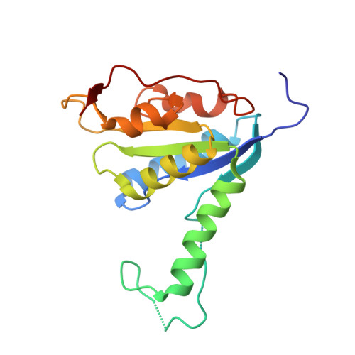
Structures of purine 2'-deoxyribosyltransferase, substrate complexes, and the ribosylated enzyme intermediate at 2.0 A resolution.
Anand, R., Kaminski, P.A., Ealick, S.E.(2004) Biochemistry 43: 2384-2393
- PubMed: 14992575
- DOI: https://doi.org/10.1021/bi035723k
- Primary Citation of Related Structures:
1S2D, 1S2G, 1S2I, 1S2L, 1S3F - PubMed Abstract:
The structure of class I N-deoxyribosyltransferase from Lactobacillus helveticus was determined by X-ray crystallography. Unlike class II N-deoxyribosyltransferases, which accept either purine or pyrimidine deoxynucleosides, class I enzymes are specific for purines as both the donor and acceptor base. Both class I and class II enzymes are highly specific for deoxynucleosides. The class I structure reveals similarities with the previously determined class II enzyme from Lactobacillus leichmanni [Armstrong, S. A., Cook, W. J., Short, S. A., and Ealick, S. E. (1996) Structure 4, 97-107]. The specificity of the class I enzyme for purine deoxynucleosides can be traced to a loop (residues 48-62), which shields the active site in the class II enzyme. In the class I enzyme, the purine base itself shields the active site from the solvent, while the smaller pyrimidine base cannot. The structure of the enzyme with a bound ribonucleoside shows that the nucleophilic oxygen atom of Glu101 hydrogen bonds to the O2' atom, rendering it unreactive and thus explaining the specificity for 2'-deoxynucleosides. The structure of a ribosylated enzyme intermediate reveals movements that occur during cleavage of the N-glycosidic bond. The structures of complexes with substrates and substrate analogues show that the purine base can bind in several different orientations, thus explaining the ability of the enzyme to catalyze alternate deoxyribosylation at the N3 or N7 position.
Organizational Affiliation:
Department of Chemistry and Chemical Biology, Cornell University, Ithaca, New York 14853, USA.


