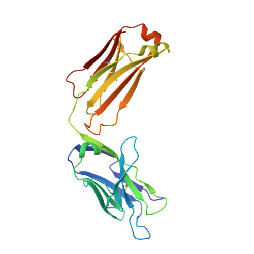
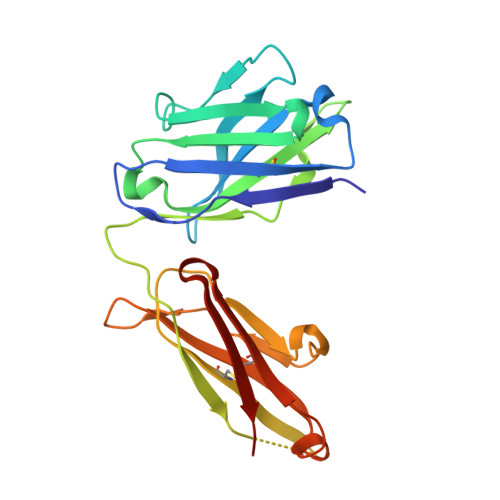
1S3K

Crystal Structure of a Humanized Fab (hu3S193) in Complex with the Lewis Y Tetrasaccharide


Experimental Data Snapshot

  • Method: X-RAY DIFFRACTION
  • Resolution: 1.90 Å
  • R-Value Free: 0.238 
  • R-Value Work: 0.203 
  • R-Value Observed: 0.203 

Starting Model: experimental
View more details

wwPDB Validation   3D Report Full Report


This is version 2.2 of the entry. See complete history


Literature

Structural convergence of antibody binding of carbohydrate determinants in lewis y tumor antigens

Ramsland, P.A.Farrugia, W.Bradford, T.M.Hogarth, P.M.Scott, A.M.

(2004) J Mol Biol 340: 809-818

  • DOI: https://doi.org/10.1016/j.jmb.2004.05.037
  • Primary Citation of Related Structures:  
    1S3K

  • PubMed Abstract: 

    Antibodies targeting human epithelial carcinomas bearing Lewis Y (Le(y)) carbohydrate antigens provide a striking illustration of convergent immune recognition. We report a 1.9A resolution crystal structure of the Fab of a humanized antibody (hu3S193) in complex with the Le(y) tetrasaccharide, Fuc(alpha 1-->2)Gal(beta 1-->4)[Fuc(alpha 1-->3)]GlcNAc. Comparisons of the hu3S193 and BR96 antibodies bound to Le(y) tumor antigens revealed extremely similar mechanisms for recognition of the carbohydrate epitopes. Solvent plays a critical role in hu3S193 antibody binding to the Le(y) carbohydrate epitope. Specificity for Le(y) is maintained because a conserved pocket accepts an N-acetyl group of the core Gal(beta 1-->4)GlcNAc disaccharide. Closely related blood-group determinants (Le(a) and Le(b)) cannot enter the specificity pocket, making the Le(y) antibodies promising candidates for immunotherapy of epithelial cancer.


  • Organizational Affiliation

    Structural Immunology Laboratory, Austin Research Institute, Heidelberg, Vic. 3084, Australia. [email protected]


Macromolecules
Find similar proteins by:  (by identity cutoff)  |  3D Structure
Entity ID: 1
MoleculeChains Sequence LengthOrganismDetailsImage
HU3S193 Fab fragment, light chainA [auth L]219Mus musculusMutation(s): 0 
Entity Groups  
Sequence Clusters30% Identity50% Identity70% Identity90% Identity95% Identity100% Identity
Sequence Annotations
Expand
  • Reference Sequence
Find similar proteins by:  (by identity cutoff)  |  3D Structure
Entity ID: 2
MoleculeChains Sequence LengthOrganismDetailsImage
HU3S193 Fab fragment, heavy chainB [auth H]222Mus musculusMutation(s): 0 
Entity Groups  
Sequence Clusters30% Identity50% Identity70% Identity90% Identity95% Identity100% Identity
Sequence Annotations
Expand
  • Reference Sequence
Oligosaccharides

Help

Entity ID: 3
MoleculeChains Length2D Diagram Glycosylation3D Interactions
alpha-L-fucopyranose-(1-2)-beta-D-galactopyranose-(1-4)-[alpha-L-fucopyranose-(1-3)]2-acetamido-2-deoxy-alpha-D-glucopyranoseC [auth A]4N/A
Glycosylation Resources
GlyTouCan:  G82236PZ
GlyCosmos:  G82236PZ
GlyGen:  G82236PZ
Small Molecules
Ligands 1 Unique
IDChains Name / Formula / InChI Key2D Diagram3D Interactions
GOL
Query on GOL

Download Ideal Coordinates CCD File 
D [auth L]GLYCEROL
C3 H8 O3
PEDCQBHIVMGVHV-UHFFFAOYSA-N
Biologically Interesting Molecules (External Reference) 1 Unique
Experimental Data & Validation

Experimental Data

  • Method: X-RAY DIFFRACTION
  • Resolution: 1.90 Å
  • R-Value Free: 0.238 
  • R-Value Work: 0.203 
  • R-Value Observed: 0.203 
  • Space Group: P 31
Unit Cell:
Length ( Å )Angle ( ˚ )
a = 71.758α = 90
b = 71.758β = 90
c = 90.142γ = 120
Software Package:
Software NamePurpose
DENZOdata reduction
SCALEPACKdata scaling
AMoREphasing
CNSrefinement

Structure Validation

View Full Validation Report


Entry History 

Deposition Data

Revision History  (Full details and data files)

  • Version 1.0: 2004-07-13
    Type: Initial release
  • Version 1.1: 2008-04-29
    Changes: Version format compliance
  • Version 1.2: 2011-07-13
    Changes: Non-polymer description, Version format compliance
  • Version 1.3: 2020-01-01
    Changes: Data collection, Derived calculations, Source and taxonomy
  • Version 2.0: 2020-07-29
    Type: Remediation
    Reason: Carbohydrate remediation
    Changes: Atomic model, Data collection, Derived calculations, Structure summary
  • Version 2.1: 2023-10-25
    Changes: Data collection, Database references, Refinement description, Structure summary
  • Version 2.2: 2024-10-30
    Changes: Structure summary