1TF0

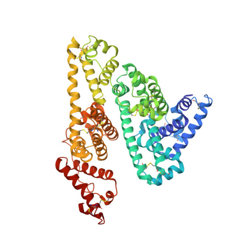
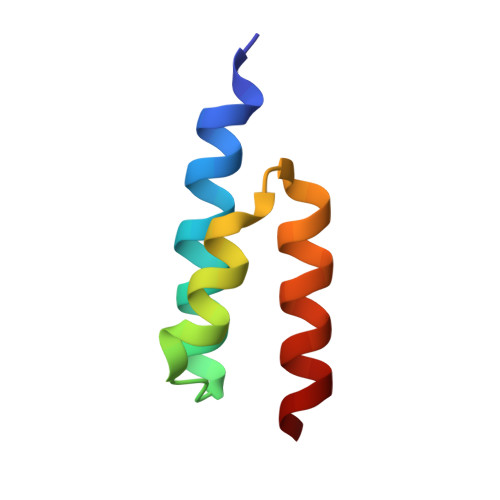
Crystal structure of the GA module complexed with human serum albumin


Experimental Data Snapshot

  • Method: X-RAY DIFFRACTION
  • Resolution: 2.70 Å
  • R-Value Free: 0.295 
  • R-Value Work: 0.249 
  • R-Value Observed: 0.251 

Starting Models: experimental
View more details

wwPDB Validation   3D Report Full Report


Ligand Structure Quality Assessment 


This is version 1.5 of the entry. See complete history


Literature

Crystal structure and biological implications of a bacterial albumin binding module in complex with human serum albumin

Lejon, S.Frick, I.-M.Bjorck, L.Wikstrom, M.Svensson, S.

(2004) J Biol Chem 279: 42924-42928

  • DOI: https://doi.org/10.1074/jbc.M406957200
  • Primary Citation of Related Structures:  
    1TF0

  • PubMed Abstract: 

    Many bactericide species express surface proteins that interact with human serum albumin (HSA). Protein PAB from the anaerobic bacterium Finegoldia magna (formerly Peptostreptococcus magnus) represents one of these proteins. Protein PAB contains a domain of 53 amino acid residues known as the GA module. GA homologs are also found in protein G of group C and G streptococci. Here we report the crystal structure of HSA in complex with the GA module of protein PAB. The model of the complex was refined to a resolution of 2.7 A and reveals a novel binding epitope located in domain II of the albumin molecule. The GA module is composed of a left-handed three-helix bundle, and residues from the second helix and the loops surrounding it were found to be involved in HSA binding. Furthermore, the presence of HSA-bound fatty acids seems to influence HSA-GA complex formation. F. magna has a much more restricted host specificity compared with C and G streptococci, which is also reflected in the binding of different animal albumins by proteins PAB and G. The structure of the HSA-GA complex offers a molecular explanation to this unusually clear example of bacterial adaptation.


  • Organizational Affiliation

    Department of Cell and Molecular Biology, Uppsala University, SE-751 24 Uppsala, Sweden. [email protected]


Macromolecules
Find similar proteins by:  (by identity cutoff)  |  3D Structure
Entity ID: 1
MoleculeChains Sequence LengthOrganismDetailsImage
Serum albumin572Homo sapiensMutation(s): 0 
UniProt & NIH Common Fund Data Resources
Find proteins for P02768 (Homo sapiens)
Explore P02768 
Go to UniProtKB:  P02768
PHAROS:  P02768
GTEx:  ENSG00000163631 
Entity Groups  
Sequence Clusters30% Identity50% Identity70% Identity90% Identity95% Identity100% Identity
UniProt GroupP02768
Sequence Annotations
Expand
  • Reference Sequence
Find similar proteins by:  (by identity cutoff)  |  3D Structure
Entity ID: 2
MoleculeChains Sequence LengthOrganismDetailsImage
Peptostreptococcal albumin-binding protein53Finegoldia magna ATCC 29328Mutation(s): 0 
Gene Names: PAB
UniProt
Find proteins for Q51911 (Finegoldia magna)
Explore Q51911 
Go to UniProtKB:  Q51911
Entity Groups  
Sequence Clusters30% Identity50% Identity70% Identity90% Identity95% Identity100% Identity
UniProt GroupQ51911
Sequence Annotations
Expand
  • Reference Sequence
Experimental Data & Validation

Experimental Data

  • Method: X-RAY DIFFRACTION
  • Resolution: 2.70 Å
  • R-Value Free: 0.295 
  • R-Value Work: 0.249 
  • R-Value Observed: 0.251 
  • Space Group: C 2 2 21
Unit Cell:
Length ( Å )Angle ( ˚ )
a = 96.295α = 90
b = 134.799β = 90
c = 122.463γ = 90
Software Package:
Software NamePurpose
REFMACrefinement
ProDCdata collection
SCALEPACKdata scaling
MOLREPphasing

Structure Validation

View Full Validation Report


Ligand Structure Quality Assessment 


Entry History 

Deposition Data

Revision History  (Full details and data files)

  • Version 1.0: 2004-09-07
    Type: Initial release
  • Version 1.1: 2008-04-30
    Changes: Version format compliance
  • Version 1.2: 2011-07-13
    Changes: Advisory, Source and taxonomy, Version format compliance
  • Version 1.3: 2017-10-11
    Changes: Refinement description
  • Version 1.4: 2023-08-23
    Changes: Data collection, Database references, Derived calculations, Refinement description
  • Version 1.5: 2024-10-09
    Changes: Structure summary