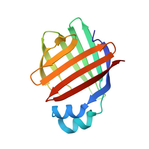
1TW4

Crystal Structure of Chicken Liver Basic Fatty Acid Binding Protein (Bile Acid Binding Protein) Complexed With Cholic Acid


Experimental Data Snapshot

  • Method: X-RAY DIFFRACTION
  • Resolution: 2.00 Å
  • R-Value Free: 0.257 
  • R-Value Work: 0.216 
  • R-Value Observed: 0.216 

wwPDB Validation   3D Report Full Report


Ligand Structure Quality Assessment 


This is version 1.4 of the entry. See complete history


Literature

Crystal structure of chicken liver basic Fatty Acid-binding protein complexed with cholic acid

Nichesola, D.Perduca, M.Capaldi, S.Carrizo, M.E.Righetti, P.G.Monaco, H.L.

(2004) Biochemistry 43: 14072-14079

  • DOI: https://doi.org/10.1021/bi0489661
  • Primary Citation of Related Structures:  
    1TVQ, 1TW4

  • PubMed Abstract: 

    Two paralogous groups of liver fatty acid-binding proteins (FABPs) have been described: the mammalian type liver FABPs and the basic type (Lb-FABPs) characterized in several vertebrates but not in mammals. The two groups have similar sequences and share a highly conserved three-dimensional structure, but their specificity and stoichiometry of binding are different. The crystal structure of chicken Lb-FABP complexed with cholic acid and that of the apoprotein refined to 2.0 A resolution are presented in this paper. The two forms of the protein crystallize in different space groups, and significant changes are observed between the two conformations. The holoprotein binds two molecules of cholate in the interior cavity, and the contacts observed between the two ligands can help to explain the reason for this stoichiometry of binding. Most of the amino acids involved in ligand binding are conserved in other members of the Lb-FABP family. Since the amino acid sequence of the Lb-FABPs is more similar to that of the bile acid-binding proteins than to that of the L-FABPs, the possibility that the Lb-FABPs might be more appropriately called liver bile acid-binding proteins (L-BABPs) is suggested.


  • Organizational Affiliation

    Laboratorio di Biocristallografía, Dipartimento Scientifico e Tecnologico, Università di Verona, Strada Le Grazie 15, 37134 Verona, Italy.


Macromolecules
Find similar proteins by:  (by identity cutoff)  |  3D Structure
Entity ID: 1
MoleculeChains Sequence LengthOrganismDetailsImage
Fatty acid-binding protein
A, B
125Gallus gallusMutation(s): 0 
UniProt
Find proteins for P80226 (Gallus gallus)
Explore P80226 
Go to UniProtKB:  P80226
Entity Groups  
Sequence Clusters30% Identity50% Identity70% Identity90% Identity95% Identity100% Identity
UniProt GroupP80226
Sequence Annotations
Expand
  • Reference Sequence
Experimental Data & Validation

Experimental Data

  • Method: X-RAY DIFFRACTION
  • Resolution: 2.00 Å
  • R-Value Free: 0.257 
  • R-Value Work: 0.216 
  • R-Value Observed: 0.216 
  • Space Group: P 21 21 21
Unit Cell:
Length ( Å )Angle ( ˚ )
a = 60.31α = 90
b = 63.41β = 90
c = 77.23γ = 90
Software Package:
Software NamePurpose
CNSrefinement
MAR345data collection
CCP4data scaling
AMoREphasing

Structure Validation

View Full Validation Report


Ligand Structure Quality Assessment 


Entry History 

Deposition Data

Revision History  (Full details and data files)

  • Version 1.0: 2004-11-23
    Type: Initial release
  • Version 1.1: 2008-04-30
    Changes: Version format compliance
  • Version 1.2: 2011-07-13
    Changes: Version format compliance
  • Version 1.3: 2017-10-11
    Changes: Refinement description
  • Version 1.4: 2024-03-13
    Changes: Data collection, Database references, Derived calculations