1U1E

Structure of e. coli uridine phosphorylase complexed to 5(phenylseleno)acyclouridine (PSAU)


Experimental Data Snapshot

  • Method: X-RAY DIFFRACTION
  • Resolution: 2.00 Å
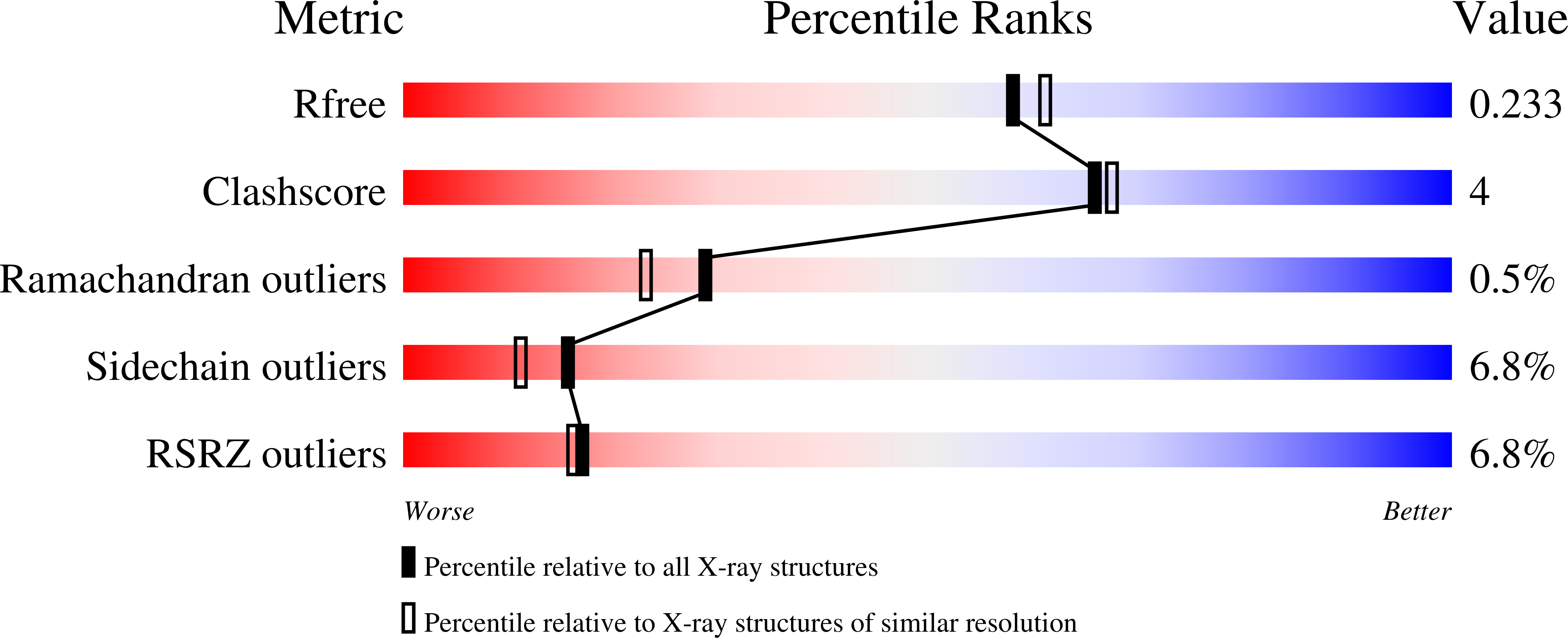
  • R-Value Free: 0.235 
  • R-Value Work: 0.211 
  • R-Value Observed: 0.214 

wwPDB Validation   3D Report Full Report


Ligand Structure Quality Assessment 


This is version 1.4 of the entry. See complete history


Literature

Structural basis for inhibition of Escherichia coli uridine phosphorylase by 5-substituted acyclouridines.

Bu, W.Settembre, E.C.el Kouni, M.H.Ealick, S.E.

(2005) Acta Crystallogr D Biol Crystallogr 61: 863-872

  • DOI: https://doi.org/10.1107/S0907444905007882
  • Primary Citation of Related Structures:  
    1U1C, 1U1D, 1U1E, 1U1F, 1U1G

  • PubMed Abstract: 

    Uridine phosphorylase (UP) catalyzes the reversible phosphorolysis of uridine to uracil and ribose 1-phosphate and is a key enzyme in the pyrimidine-salvage pathway. Escherichia coli UP is structurally homologous to E. coli purine nucleoside phosphorylase and other members of the type I family of nucleoside phosphorylases. The structures of 5-benzylacyclouridine, 5-phenylthioacyclouridine, 5-phenylselenenylacyclouridine, 5-m-benzyloxybenzyl acyclouridine and 5-m-benzyloxybenzyl barbituric acid acyclonucleoside bound to the active site of E. coli UP have been determined, with resolutions ranging from 1.95 to 2.3 A. For all five complexes the acyclo sugar moiety binds to the active site in a conformation that mimics the ribose ring of the natural substrates. Surprisingly, the terminal hydroxyl group occupies the position of the nonessential 5'-hydroxyl substituent of the substrate rather than the 3'-hydroxyl group, which is normally required for catalytic activity. Until recently, inhibitors of UP were designed with limited structural knowledge of the active-site residues. These structures explain the basis of inhibition for this series of acyclouridine analogs and suggest possible additional avenues for future drug-design efforts. Furthermore, the studies can be extended to design inhibitors of human UP, for which no X-ray structure is available.


  • Organizational Affiliation

    Department of Chemistry and Chemical Biology, Cornell University, Ithaca, NY 14853-1301, USA.


Macromolecules
Find similar proteins by:  (by identity cutoff)  |  3D Structure
Entity ID: 1
MoleculeChains Sequence LengthOrganismDetailsImage
Uridine phosphorylase
A, B, C, D, E
A, B, C, D, E, F
256Escherichia coliMutation(s): 0 
Gene Names: UDPB3831
EC: 2.4.2.3
UniProt
Find proteins for P12758 (Escherichia coli (strain K12))
Explore P12758 
Go to UniProtKB:  P12758
Entity Groups  
Sequence Clusters30% Identity50% Identity70% Identity90% Identity95% Identity100% Identity
UniProt GroupP12758
Sequence Annotations
Expand
  • Reference Sequence
Small Molecules
Ligands 3 Unique
IDChains Name / Formula / InChI Key2D Diagram3D Interactions
182
Query on 182

Download Ideal Coordinates CCD File 
I [auth A]
K [auth B]
N [auth C]
P [auth D]
S [auth E]
I [auth A],
K [auth B],
N [auth C],
P [auth D],
S [auth E],
U [auth F]
1-((2-HYDROXYETHOXY)METHYL)-5-(PHENYLSELANYL)PYRIMIDINE-2,4(1H,3H)-DIONE
C13 H14 N2 O4 Se
KFSPSGWABMNFIY-UHFFFAOYSA-N
PO4
Query on PO4

Download Ideal Coordinates CCD File 
G [auth A]
J [auth B]
L [auth C]
O [auth D]
Q [auth E]
G [auth A],
J [auth B],
L [auth C],
O [auth D],
Q [auth E],
T [auth F]
PHOSPHATE ION
O4 P
NBIIXXVUZAFLBC-UHFFFAOYSA-K
K
Query on K

Download Ideal Coordinates CCD File 
H [auth A],
M [auth C],
R [auth E]
POTASSIUM ION
K
NPYPAHLBTDXSSS-UHFFFAOYSA-N
Experimental Data & Validation

Experimental Data

  • Method: X-RAY DIFFRACTION
  • Resolution: 2.00 Å
  • R-Value Free: 0.235 
  • R-Value Work: 0.211 
  • R-Value Observed: 0.214 
  • Space Group: P 21 21 21
Unit Cell:
Length ( Å )Angle ( ˚ )
a = 91.076α = 90
b = 125.69β = 90
c = 140.899γ = 90
Software Package:
Software NamePurpose
REFMACrefinement
SCALEPACKdata scaling
CNSrefinement
MOSFLMdata reduction
CNSphasing

Structure Validation

View Full Validation Report


Ligand Structure Quality Assessment 


Entry History 

Deposition Data

Revision History  (Full details and data files)

  • Version 1.0: 2005-07-05
    Type: Initial release
  • Version 1.1: 2008-04-30
    Changes: Version format compliance
  • Version 1.2: 2011-07-13
    Changes: Advisory, Version format compliance
  • Version 1.3: 2019-07-24
    Changes: Data collection, Refinement description
  • Version 1.4: 2024-02-14
    Changes: Data collection, Database references, Derived calculations, Refinement description