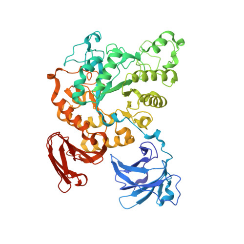
1VFO

Crystal structure of Thermoactinomyces vulgaris R-47 alpha-amylase 2/beta-cyclodextrin complex


Experimental Data Snapshot

  • Method: X-RAY DIFFRACTION
  • Resolution: 2.81 Å
  • R-Value Free: 0.256 
  • R-Value Work: 0.200 
  • R-Value Observed: 0.200 

wwPDB Validation   3D Report Full Report


This is version 2.2 of the entry. See complete history


Literature

Complex structures of Thermoactinomyces vulgaris R-47 alpha-amylase 2 with acarbose and cyclodextrins demonstrate the multiple substrate recognition mechanism

Ohtaki, A.Mizuno, M.Tonozuka, T.Sakano, Y.Kamitori, S.

(2004) J Biol Chem 279: 31033-31040

  • DOI: https://doi.org/10.1074/jbc.M404311200
  • Primary Citation of Related Structures:  
    1VFM, 1VFO, 1VFU, 3A6O

  • PubMed Abstract: 

    Thermoactinomyces vulgaris R-47 alpha-amylase 2 (TVAII) has the unique ability to hydrolyze cyclodextrins (CDs), with various sized cavities, as well as starch. To understand the relationship between structure and substrate specificity, x-ray structures of a TVAII-acarbose complex and inactive mutant TVAII (D325N/D421N)/alpha-, beta- and gamma-CDs complexes were determined at resolutions of 2.9, 2.9, 2.8, and 3.1 A, respectively. In all complexes, the interactions between ligands and enzymes at subsites -1, -2, and -3 were almost the same, but striking differences in the catalytic site structure were found at subsites +1 and +2, where Trp(356) and Tyr(374) changed the conformation of the side chain depending on the structure and size of the ligands. Trp(356) and Tyr(374) are thought to be responsible for the multiple substrate-recognition mechanism of TVAII, providing the unique substrate specificity. In the beta-CD complex, the beta-CD maintains a regular conical structure, making it difficult for Glu(354) to protonate the O-4 atom at the hydrolyzing site as a previously proposed hydrolyzing mechanism of alpha-amylase. From the x-ray structures, it is suggested that the protonation of the O-4 atom is possibly carried out via a hydrogen atom of the inter-glucose hydrogen bond at the hydrolyzing site.


  • Organizational Affiliation

    Department of Biotechnology and Life Science, Tokyo University of Agriculture and Technology, 2-24-16 Naka-cho, Koganei, Tokyo 184-8588, Japan.


Macromolecules
Find similar proteins by:  (by identity cutoff)  |  3D Structure
Entity ID: 1
MoleculeChains Sequence LengthOrganismDetailsImage
Neopullulanase 2
A, B
585Thermoactinomyces vulgarisMutation(s): 2 
EC: 3.2.1.135
UniProt
Find proteins for Q08751 (Thermoactinomyces vulgaris)
Explore Q08751 
Go to UniProtKB:  Q08751
Entity Groups  
Sequence Clusters30% Identity50% Identity70% Identity90% Identity95% Identity100% Identity
UniProt GroupQ08751
Sequence Annotations
Expand
  • Reference Sequence
Oligosaccharides

Help

Entity ID: 2
MoleculeChains Length2D Diagram Glycosylation3D Interactions
Cycloheptakis-(1-4)-(alpha-D-glucopyranose)
C
7N/A
Glycosylation Resources
GlyTouCan:  G01435GL
GlyCosmos:  G01435GL
Entity ID: 3
MoleculeChains Length2D Diagram Glycosylation3D Interactions
Cyclic alpha-D-glucopyranose-(1-4)-beta-D-glucopyranose-(1-4)-alpha-D-glucopyranose-(1-4)-alpha-D-glucopyranose-(1-4)-alpha-D-glucopyranose-(1-4)-alpha-D-glucopyranose-(1-4)-alpha-D-glucopyranose
D
7N/A
Glycosylation Resources
GlyTouCan:  G86771JB
GlyCosmos:  G86771JB
Biologically Interesting Molecules (External Reference) 1 Unique
Experimental Data & Validation

Experimental Data

  • Method: X-RAY DIFFRACTION
  • Resolution: 2.81 Å
  • R-Value Free: 0.256 
  • R-Value Work: 0.200 
  • R-Value Observed: 0.200 
  • Space Group: P 21 21 21
Unit Cell:
Length ( Å )Angle ( ˚ )
a = 115.083α = 90
b = 118.812β = 90
c = 114.277γ = 90
Software Package:
Software NamePurpose
CNSrefinement
MOSFLMdata reduction
CCP4data scaling
CNSphasing

Structure Validation

View Full Validation Report


Entry History 

Deposition Data

Revision History  (Full details and data files)

  • Version 1.0: 2005-02-08
    Type: Initial release
  • Version 1.1: 2008-04-27
    Changes: Version format compliance
  • Version 1.2: 2011-07-13
    Changes: Version format compliance
  • Version 2.0: 2020-07-29
    Type: Remediation
    Reason: Carbohydrate remediation
    Changes: Atomic model, Data collection, Derived calculations, Structure summary
  • Version 2.1: 2021-11-10
    Changes: Database references, Structure summary
  • Version 2.2: 2023-12-27
    Changes: Data collection