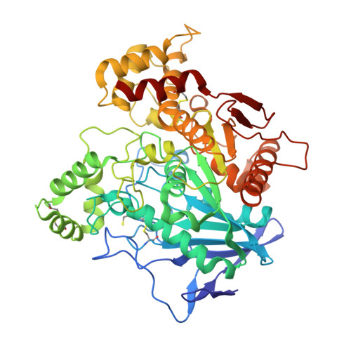
1XLV

Ethylphosphorylated Butyrylcholinesterase (Aged) Obtained By Reaction With Echothiophate


Experimental Data Snapshot

  • Method: X-RAY DIFFRACTION
  • Resolution: 2.25 Å
  • R-Value Free: 0.247 
  • R-Value Work: 0.183 
  • R-Value Observed: 0.186 

Starting Model: experimental
View more details

wwPDB Validation   3D Report Full Report


Ligand Structure Quality Assessment 


This is version 2.4 of the entry. See complete history


Literature

Role of Water in Aging of Human Butyrylcholinesterase Inhibited by Echothiophate: The Crystal Structure Suggests Two Alternative Mechanisms of Aging

Nachon, F.Asojo, O.A.Borgstahl, G.E.O.Masson, P.Lockridge, O.

(2005) Biochemistry 44: 1154-1162

  • DOI: https://doi.org/10.1021/bi048238d
  • Primary Citation of Related Structures:  
    1XLU, 1XLV, 1XLW

  • PubMed Abstract: 

    Organophosphorus poisons (OP) bind covalently to the active-site serine of cholinesterases. The inhibited enzyme can usually be reactivated with powerful nucleophiles such as oximes. However, the covalently bound OP can undergo a suicide reaction (termed aging) yielding nonreactivatable enzyme. In human butyrylcholinesterase (hBChE), aging involves the residues His438 and Glu197 that are proximal to the active-site serine (Ser198). The mechanism of aging is known in detail for the nerve gases soman, sarin, and tabun as well as the pesticide metabolite isomalathion. Aging of soman- and sarin-inhibited acetylcholinesterase occurs by C-O bond cleavage, whereas that of tabun- and isomalathion-inhibited acetylcholinesterase occurs by P-N and P-S bond cleavage, respectively. In this work, the crystal structures of hBChE inhibited by the ophthalmic reagents echothiophate (nonaged and aged) and diisopropylfluorophosphate (aged) were solved and refined to 2.1, 2.25, and 2.2 A resolution, respectively. No appreciable shift in the position of the catalytic triad histidine was observed between the aged and nonaged conjugates of hBChE. This absence of shift contrasts with the aged and nonaged crystal structures of Torpedo californica acetylcholinesterase inhibited by the nerve agent VX. The nonaged hBChE structure shows one water molecule interacting with Glu197 and the catalytic triad histidine (His438). Interestingly, this water molecule is ideally positioned to promote aging by two mechanisms: breaking either a C-O bond or a P-O bond. Pesticides and certain stereoisomers of nerve agents are expected to undergo aging by breaking the P-O bond.


  • Organizational Affiliation

    The Eppley Institute for Research in Cancer and Allied Diseases, University of Nebraska Medical Center, Omaha, Nebraska 68198-6805, USA. [email protected]


Macromolecules
Find similar proteins by:  (by identity cutoff)  |  3D Structure
Entity ID: 1
MoleculeChains Sequence LengthOrganismDetailsImage
BUTYRYLCHOLINESTERASE529Homo sapiensMutation(s): 4 
EC: 3.1.1.8
UniProt & NIH Common Fund Data Resources
Find proteins for P06276 (Homo sapiens)
Explore P06276 
Go to UniProtKB:  P06276
PHAROS:  P06276
GTEx:  ENSG00000114200 
Entity Groups  
Sequence Clusters30% Identity50% Identity70% Identity90% Identity95% Identity100% Identity
UniProt GroupP06276
Glycosylation
Glycosylation Sites: 6Go to GlyGen: P06276-1
Sequence Annotations
Expand
  • Reference Sequence
Oligosaccharides

Help

Entity ID: 2
MoleculeChains Length2D Diagram Glycosylation3D Interactions
2-acetamido-2-deoxy-beta-D-glucopyranose-(1-4)-[beta-L-fucopyranose-(1-6)]2-acetamido-2-deoxy-beta-D-glucopyranose
B
3N-Glycosylation
Glycosylation Resources
GlyTouCan:  G28454KX
GlyCosmos:  G28454KX
GlyGen:  G28454KX
Entity ID: 3
MoleculeChains Length2D Diagram Glycosylation3D Interactions
2-acetamido-2-deoxy-beta-D-glucopyranose-(1-4)-2-acetamido-2-deoxy-beta-D-glucopyranose
C
2N-Glycosylation
Glycosylation Resources
GlyTouCan:  G42666HT
GlyCosmos:  G42666HT
GlyGen:  G42666HT
Small Molecules
Ligands 5 Unique
IDChains Name / Formula / InChI Key2D Diagram3D Interactions
NAG
Query on NAG

Download Ideal Coordinates CCD File 
D [auth A],
E [auth A],
F [auth A],
G [auth A]
2-acetamido-2-deoxy-beta-D-glucopyranose
C8 H15 N O6
OVRNDRQMDRJTHS-FMDGEEDCSA-N
EFS
Query on EFS

Download Ideal Coordinates CCD File 
N [auth A]ETHYL DIHYDROGEN PHOSPHATE
C2 H7 O4 P
ZJXZSIYSNXKHEA-UHFFFAOYSA-N
SO4
Query on SO4

Download Ideal Coordinates CCD File 
H [auth A],
I [auth A],
J [auth A]
SULFATE ION
O4 S
QAOWNCQODCNURD-UHFFFAOYSA-L
GOL
Query on GOL

Download Ideal Coordinates CCD File 
O [auth A],
P [auth A],
Q [auth A],
R [auth A]
GLYCEROL
C3 H8 O3
PEDCQBHIVMGVHV-UHFFFAOYSA-N
CL
Query on CL

Download Ideal Coordinates CCD File 
K [auth A],
L [auth A],
M [auth A]
CHLORIDE ION
Cl
VEXZGXHMUGYJMC-UHFFFAOYSA-M
Modified Residues  1 Unique
IDChains TypeFormula2D DiagramParent
CSS
Query on CSS
A
L-PEPTIDE LINKINGC3 H7 N O2 S2CYS
Experimental Data & Validation

Experimental Data

  • Method: X-RAY DIFFRACTION
  • Resolution: 2.25 Å
  • R-Value Free: 0.247 
  • R-Value Work: 0.183 
  • R-Value Observed: 0.186 
  • Space Group: I 4 2 2
Unit Cell:
Length ( Å )Angle ( ˚ )
a = 154.587α = 90
b = 154.587β = 90
c = 126.706γ = 90
Software Package:
Software NamePurpose
SCALEPACKdata scaling
AMoREphasing
REFMACrefinement

Structure Validation

View Full Validation Report


Ligand Structure Quality Assessment 


Entry History 

Deposition Data

Revision History  (Full details and data files)

  • Version 1.0: 2005-02-01
    Type: Initial release
  • Version 1.1: 2008-04-30
    Changes: Version format compliance
  • Version 1.2: 2011-07-13
    Changes: Non-polymer description, Version format compliance
  • Version 1.3: 2017-10-11
    Changes: Refinement description
  • Version 2.0: 2020-07-29
    Type: Remediation
    Reason: Carbohydrate remediation
    Changes: Atomic model, Data collection, Derived calculations, Structure summary
  • Version 2.1: 2021-10-20
    Changes: Database references, Derived calculations, Structure summary
  • Version 2.2: 2023-08-23
    Changes: Data collection, Refinement description
  • Version 2.3: 2023-11-15
    Changes: Data collection
  • Version 2.4: 2024-10-30
    Changes: Structure summary