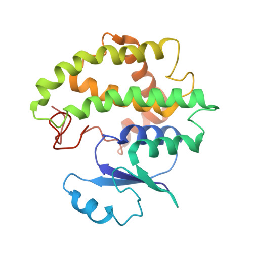
1Y6E

Orthorhombic glutathione S-transferase of Schistosoma japonicum


Experimental Data Snapshot

  • Method: X-RAY DIFFRACTION
  • Resolution: 3.00 Å
  • R-Value Free: 0.279 
  • R-Value Work: 0.211 
  • R-Value Observed: 0.215 

wwPDB Validation   3D Report Full Report


This is version 1.3 of the entry. See complete history


Literature

X-ray structure of glutathione S-transferase from Schistosoma japonicum in a new crystal form reveals flexibility of the substrate-binding site

Rufer, A.C.Thiebach, L.Baer, K.Klein, H.W.Hennig, M.

(2005) Acta Crystallogr Sect F Struct Biol Cryst Commun 61: 263-265

  • DOI: https://doi.org/10.1107/S1744309105004823
  • Primary Citation of Related Structures:  
    1Y6E

  • PubMed Abstract: 

    The crystal structure of the 26 kDa glutathione S-transferase from Schistosoma japonicum (SjGST) was determined at 3 A resolution in the new space group P2(1)2(1)2(1). The structure of orthorhombic SjGST reveals unique features of the ligand-binding site and dimer interface when compared with previously reported structures. SjGST is recognized as the major detoxification enzyme of S. japonicum, a pathogenic helminth causing schistosomiasis. As resistance against the established inhibitor of SjGST, praziquantel, has been reported these results might prove to be valuable for the development of novel drugs.


  • Organizational Affiliation

    F. Hoffmann-La Roche AG, Pharma Research Discovery Chemistry, 4070 Basel, Switzerland. [email protected]


Macromolecules
Find similar proteins by:  (by identity cutoff)  |  3D Structure
Entity ID: 1
MoleculeChains Sequence LengthOrganismDetailsImage
glutathione S-transferase
A, B
223Schistosoma japonicumMutation(s): 0 
EC: 2.5.1.18
UniProt
Find proteins for P08515 (Schistosoma japonicum)
Explore P08515 
Go to UniProtKB:  P08515
Entity Groups  
Sequence Clusters30% Identity50% Identity70% Identity90% Identity95% Identity100% Identity
UniProt GroupP08515
Sequence Annotations
Expand
  • Reference Sequence
Experimental Data & Validation

Experimental Data

  • Method: X-RAY DIFFRACTION
  • Resolution: 3.00 Å
  • R-Value Free: 0.279 
  • R-Value Work: 0.211 
  • R-Value Observed: 0.215 
  • Space Group: P 21 21 21
Unit Cell:
Length ( Å )Angle ( ˚ )
a = 161.22α = 90
b = 50.75β = 90
c = 57.5γ = 90
Software Package:
Software NamePurpose
DENZOdata reduction
SCALEPACKdata scaling
MOLREPphasing
CNSrefinement

Structure Validation

View Full Validation Report


Entry History 

Deposition Data

Revision History  (Full details and data files)

  • Version 1.0: 2005-03-08
    Type: Initial release
  • Version 1.1: 2008-04-30
    Changes: Version format compliance
  • Version 1.2: 2011-07-13
    Changes: Version format compliance
  • Version 1.3: 2024-03-13
    Changes: Data collection, Database references