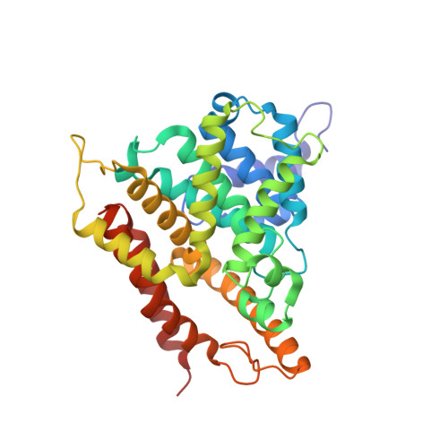
Crystal Structures of Phosphodiesterases 4 and 5 in Complex with Inhibitor 3-Isobutyl-1-Methylxanthine Suggest a Conformation Determinant of Inhibitor Selectivity
Huai, Q., Liu, Y., Francis, S.H., Corbin, J.D., Ke, H.(2004) J Biol Chem 279: 13095-13101
- PubMed: 14668322
- DOI: https://doi.org/10.1074/jbc.M311556200
- Primary Citation of Related Structures:
1RKP, 1ZKN - PubMed Abstract:
Cyclic nucleotide phosphodiesterases (PDEs) are a superfamily of enzymes controlling cellular concentrations of the second messengers cAMP and cGMP. Crystal structures of the catalytic domains of cGMP-specific PDE5A1 and cAMP-specific PDE4D2 in complex with the nonselective inhibitor 3-isobutyl-1-methylxanthine have been determined at medium resolution. The catalytic domain of PDE5A1 has the same topological folding as that of PDE4D2, but three regions show different tertiary structures, including residues 79-113, 208-224 (H-loop), and 341-364 (M-loop) in PDE4D2 or 535-566, 661-676, and 787-812 in PDE5A1, respectively. Because H- and M-loops are involved in binding of the selective inhibitors, the different conformations of the loops, thus the distinct shapes of the active sites, will be a determinant of inhibitor selectivity in PDEs. IBMX binds to a subpocket that comprises key residues Ile-336, Phe-340, Gln-369, and Phe-372 of PDE4D2 or Val-782, Phe-786, Gln-817, and Phe-820 of PDE5A1. This subpocket may be a common site for binding nonselective inhibitors of PDEs.
Organizational Affiliation:
Department of Biochemistry and Biophysics and Lineberger Comprehensive Cancer Center, the University of North Carolina, Chapel Hill, NC 27599-7260, USA.


