1B02

CRYSTAL STRUCTURE OF THYMIDYLATE SYNTHASE A FROM BACILLUS SUBTILIS


Experimental Data Snapshot

  • Method: X-RAY DIFFRACTION
  • Resolution: 2.50 Å
  • R-Value Free: 0.233 
  • R-Value Work: 0.194 

Starting Model: experimental
View more details

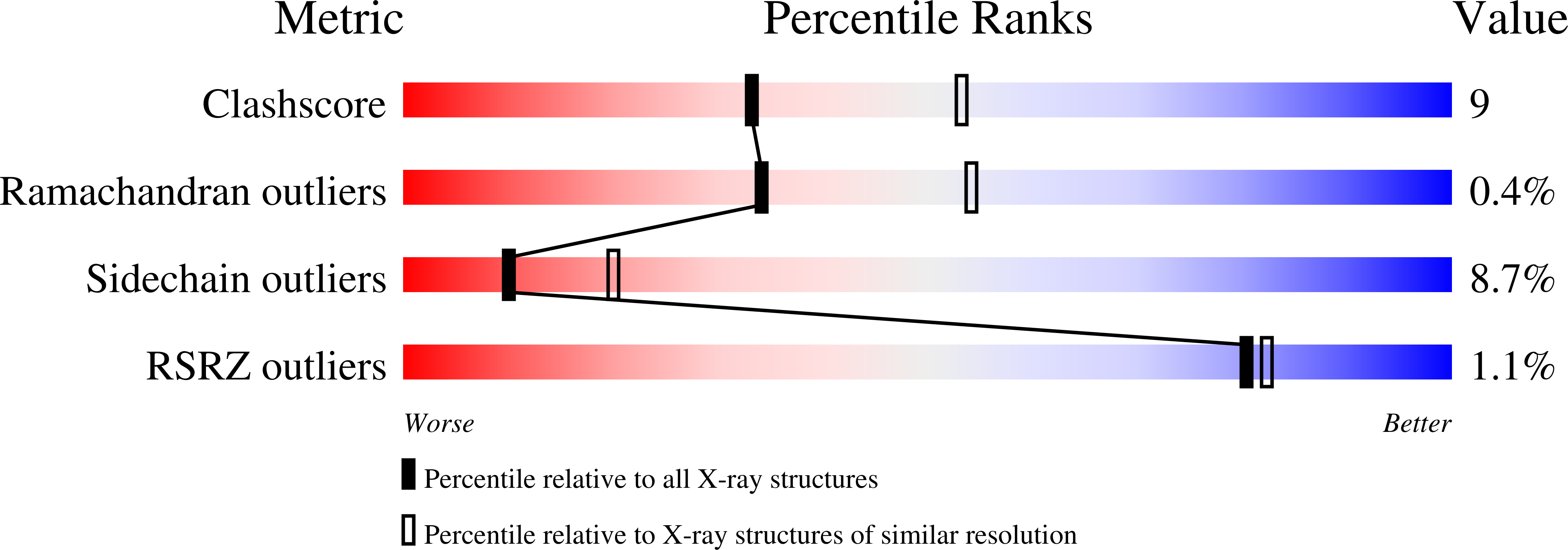
wwPDB Validation   3D Report Full Report


Ligand Structure Quality Assessment 


This is version 1.5 of the entry. See complete history


Literature

Crystal structure of thymidylate synthase A from Bacillus subtilis.

Fox, K.M.Maley, F.Garibian, A.Changchien, L.M.Van Roey, P.

(1999) Protein Sci 8: 538-544

  • DOI: https://doi.org/10.1110/ps.8.3.538
  • Primary Citation of Related Structures:  
    1B02

  • PubMed Abstract: 

    Thymidylate synthase (TS) converts dUMP to dTMP by reductive methylation, where 5,10-methylenetetrahydrofolate is the source of both the methylene group and reducing equivalents. The mechanism of this reaction has been extensively studied, mainly using the enzyme from Escherichia coli. Bacillus subtilis contains two genes for TSs, ThyA and ThyB. The ThyB enzyme is very similar to other bacterial TSs, but the ThyA enzyme is quite different, both in sequence and activity. In ThyA TS, the active site histidine is replaced by valine. In addition, the B. subtilis enzyme has a 2.4-fold greater k(cat) than the E. coli enzyme. The structure of B. subtilis thymidylate synthase in a ternary complex with 5-fluoro-dUMP and 5,10-methylenetetrahydrofolate has been determined to 2.5 A resolution. Overall, the structure of B. subtilis TS (ThyA) is similar to that of the E. coli enzyme. However, there are significant differences in the structures of two loops, the dimer interface and the details of the active site. The effects of the replacement of histidine by valine and a serine to glutamine substitution in the active site area, and the addition of a loop over the carboxy terminus may account for the differences in k(cat) found between the two enzymes.


  • Organizational Affiliation

    Department of Chemistry, Union College, Schenectady, New York 12308, USA.


Macromolecules
Find similar proteins by:  (by identity cutoff)  |  3D Structure
Entity ID: 1
MoleculeChains Sequence LengthOrganismDetailsImage
PROTEIN (THYMIDYLATE SYNTHASE)279Bacillus subtilisMutation(s): 0 
EC: 2.1.1.45
UniProt
Find proteins for P0CI79 (Bacillus subtilis (strain 168))
Explore P0CI79 
Go to UniProtKB:  P0CI79
Entity Groups  
Sequence Clusters30% Identity50% Identity70% Identity90% Identity95% Identity100% Identity
UniProt GroupP0CI79
Sequence Annotations
Expand
  • Reference Sequence
Small Molecules
Ligands 2 Unique
IDChains Name / Formula / InChI Key2D Diagram3D Interactions
C2F
Query on C2F

Download Ideal Coordinates CCD File 
C [auth A]5-METHYL-5,6,7,8-TETRAHYDROFOLIC ACID
C20 H25 N7 O6
ZNOVTXRBGFNYRX-STQMWFEESA-N
UFP
Query on UFP

Download Ideal Coordinates CCD File 
B [auth A]5-FLUORO-2'-DEOXYURIDINE-5'-MONOPHOSPHATE
C9 H12 F N2 O8 P
HFEKDTCAMMOLQP-RRKCRQDMSA-N
Experimental Data & Validation

Experimental Data

  • Method: X-RAY DIFFRACTION
  • Resolution: 2.50 Å
  • R-Value Free: 0.233 
  • R-Value Work: 0.194 
  • Space Group: P 21 21 21
Unit Cell:
Length ( Å )Angle ( ˚ )
a = 55.57α = 90
b = 105.01β = 90
c = 117.26γ = 90
Software Package:
Software NamePurpose
AMoREphasing
X-PLORrefinement
DENZOdata reduction
SCALEPACKdata scaling

Structure Validation

View Full Validation Report


Ligand Structure Quality Assessment 


Entry History 

Deposition Data

Revision History  (Full details and data files)

  • Version 1.0: 1999-03-29
    Type: Initial release
  • Version 1.1: 2008-04-26
    Changes: Version format compliance
  • Version 1.2: 2011-07-13
    Changes: Derived calculations, Version format compliance
  • Version 1.3: 2014-01-29
    Changes: Other
  • Version 1.4: 2023-08-09
    Changes: Data collection, Database references, Derived calculations, Refinement description
  • Version 1.5: 2024-11-13
    Changes: Structure summary