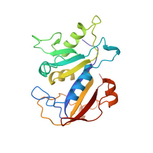
The structure of Pneumocystis carinii dihydrofolate reductase to 1.9 A resolution.
Champness, J.N., Achari, A., Ballantine, S.P., Bryant, P.K., Delves, C.J., Stammers, D.K.(1994) Structure 2: 915-924
- PubMed: 7866743
- DOI: https://doi.org/10.1016/s0969-2126(94)00093-x
- Primary Citation of Related Structures:
1DYR - PubMed Abstract:
The fungal pathogen Pneumocystis carinii causes a pneumonia which is an opportunistic infection of AIDS patients. Current therapy includes the dihydrofolate reductase (DHFR) inhibitor trimethoprim which is selective but only a relatively weak inhibitor of the enzyme for P. carinii. Determination of the three-dimensional structure of the enzyme should form the basis for design of more potent and selective therapeutic agents for treatment of the disease. The structure of P. carinii DHFR in complex with reduced nicotinamide adenine dinucleotide phosphate and trimethoprim has accordingly been solved by X-ray crystallography. The structure of the ternary complex has been refined at 1.86 A resolution (R = 0.181). A similar ternary complex with piritrexim (which is a tighter binding, but less selective inhibitor) has also been solved, as has the binary complex holoenzyme, both at 2.5 A resolution. These structures show how two drugs interact with a fungal DHFR. A comparison of the three-dimensional structure of this relatively large DHFR with bacterial or mammalian enzyme-inhibitor complexes determined previously highlights some additional secondary structure elements in this particular enzyme species. These comparisons provide further insight into the principles governing DHFR-inhibitor interaction, in which the volume of the active site appears to determine the strength of inhibitor binding.
Organizational Affiliation:
Physical Sciences Department, Wellcome Research Laboratories, Beckenham, Kent, UK.


