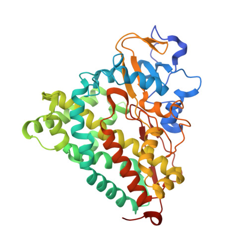
Production and X-ray crystallographic analysis of fully deuterated cytochrome P450cam.
Meilleur, F., Dauvergne, M.T., Schlichting, I., Myles, D.A.(2005) Acta Crystallogr D Biol Crystallogr 61: 539-544
- PubMed: 15858263
- DOI: https://doi.org/10.1107/S0907444905003872
- Primary Citation of Related Structures:
1YRC, 1YRD - PubMed Abstract:
Neutron protein crystallography allows H-atom positions to be located in biological structures at the relatively modest resolution of 1.5-2.0 A. A difficulty of this technique arises from the incoherent scattering from hydrogen, which considerably reduces the signal-to-noise ratio of the data. This can be overcome by preparing fully deuterated samples. Efficient protocols for routine and low-cost production of in vivo deuterium-enriched proteins have been developed. Here, the overexpression and crystallization of highly (>99%) deuterium-enriched cytochrome P450cam for neutron analysis is reported. Cytochrome P450cam from Pseudomonas putida catalyses the hydroxylation of camphor from haem-bound molecular O(2) via a mechanism that is thought to involve a proton-shuttle pathway to the active site. Since H atoms cannot be visualized in available X-ray structures, neutron diffraction is being used to determine the protonation states and water structure at the active site of the enzyme. Analysis of both hydrogenated and perdeuterated P450cam showed no significant changes between the X-ray structures determined at 1.4 and 1.7 A, respectively. This work demonstrates that the fully deuterated protein is highly isomorphous with the native (hydrogenated) protein and is appropriate for neutron protein crystallographic analysis.
Organizational Affiliation:
European Molecular Laboratory Grenoble Outstation, 6 Rue Jules Horowitz, 38042 Grenoble, France. [email protected]


