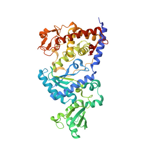
2B56

Structural Basis for UTP Specificity of RNA Editing TUTases From Trypanosoma Brucei


Experimental Data Snapshot

  • Method: X-RAY DIFFRACTION
  • Resolution: 1.97 Å
  • R-Value Free: 0.221 
  • R-Value Work: 0.193 
  • R-Value Observed: 0.194 

wwPDB Validation   3D Report Full Report


Ligand Structure Quality Assessment 


This is version 1.4 of the entry. See complete history


Literature

Structural basis for UTP specificity of RNA editing TUTases from Trypanosoma brucei.

Deng, J.Ernst, N.L.Turley, S.Stuart, K.D.Hol, W.G.

(2005) EMBO J 24: 4007-4017

  • DOI: https://doi.org/10.1038/sj.emboj.7600861
  • Primary Citation of Related Structures:  
    2B4V, 2B51, 2B56

  • PubMed Abstract: 

    Trypanosomatids are pathogenic protozoa that undergo a unique form of post-transcriptional RNA editing that inserts or deletes uridine nucleotides in many mitochondrial pre-mRNAs. Editing is catalyzed by a large multiprotein complex, the editosome. A key editosome enzyme, RNA editing terminal uridylyl transferase 2 (TUTase 2; RET2) catalyzes the uridylate addition reaction. Here, we report the 1.8 A crystal structure of the Trypanosoma brucei RET2 apoenzyme and its complexes with uridine nucleotides. This structure reveals that the specificity of the TUTase for UTP is determined by a crucial water molecule that is exquisitely positioned by the conserved carboxylates D421 and E424 to sense a hydrogen atom on the N3 position of the uridine base. The three-domain structure also unveils a unique domain arrangement not seen before in the nucleotidyltansferase superfamily, with a large domain insertion between the catalytic aspartates. This insertion is present in all trypanosomatid TUTases. We also show that TbRET2 is essential for survival of the bloodstream form of the parasite and therefore is a potential target for drug therapy.


  • Organizational Affiliation

    Howard Hughes Medical Institute, University of Washington, Seattle, WA 98195, USA.


Macromolecules
Find similar proteins by:  (by identity cutoff)  |  3D Structure
Entity ID: 1
MoleculeChains Sequence LengthOrganismDetailsImage
RNA editing complex protein MP57468Trypanosoma bruceiMutation(s): 3 
Gene Names: Tbmp57
EC: 2.7.7.52
UniProt
Find proteins for Q86MV5 (Trypanosoma brucei brucei)
Explore Q86MV5 
Go to UniProtKB:  Q86MV5
Entity Groups  
Sequence Clusters30% Identity50% Identity70% Identity90% Identity95% Identity100% Identity
UniProt GroupQ86MV5
Sequence Annotations
Expand
  • Reference Sequence
Experimental Data & Validation

Experimental Data

  • Method: X-RAY DIFFRACTION
  • Resolution: 1.97 Å
  • R-Value Free: 0.221 
  • R-Value Work: 0.193 
  • R-Value Observed: 0.194 
  • Space Group: P 41 21 2
Unit Cell:
Length ( Å )Angle ( ˚ )
a = 91.435α = 90
b = 91.435β = 90
c = 162.529γ = 90
Software Package:
Software NamePurpose
REFMACrefinement
HKL-2000data reduction
SCALEPACKdata scaling

Structure Validation

View Full Validation Report


Ligand Structure Quality Assessment 


Entry History 

Deposition Data

Revision History  (Full details and data files)

  • Version 1.0: 2005-11-22
    Type: Initial release
  • Version 1.1: 2008-05-01
    Changes: Version format compliance
  • Version 1.2: 2011-07-13
    Changes: Advisory, Version format compliance
  • Version 1.3: 2021-10-20
    Changes: Database references, Derived calculations
  • Version 1.4: 2024-02-14
    Changes: Data collection