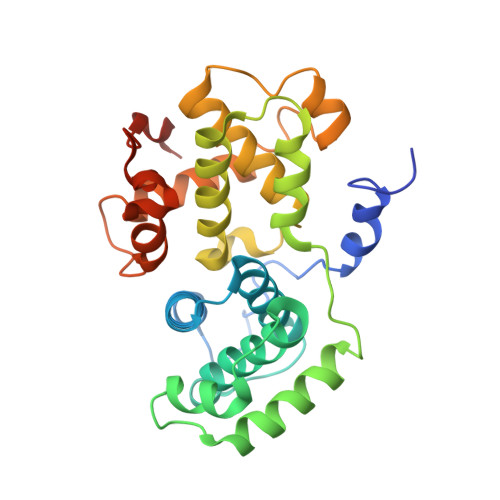
The Crystal Structure of Human Cyclin B.
Petri, E.T., Errico, A., Escobedo, L., Hunt, T., Basavappa, R.(2007) Cell Cycle 6: 1342-1349
- PubMed: 17495533
- DOI: https://doi.org/10.4161/cc.6.11.4297
- Primary Citation of Related Structures:
2B9R - PubMed Abstract:
Cyclin B is the key regulatory protein controlling mitosis in all eukaryotes, where it binds cyclin-dependent kinase, cdk1, forming a complex which initiates the mitotic program through phosphorylation of select proteins. Cyclin B regulates the activation, subcellular localization, and substrate specificity of cdk1, and destruction of cyclin B is necessary for mitotic exit. Overexpression of human cyclin B1 has been found in numerous cancers and has been associated with tumor aggressiveness. Here we report the crystal structure of human cyclin B1 to 2.9 A. Comparison of the structure with cyclin A and cyclin E reveals remarkably similar N-terminal cyclin box motifs but significant differences among the C-terminal cyclin box lobes. Divergence in sequence gives rise to unique interaction surfaces at the proposed cyclin B/cdk1 interface as well as the 'RxL' motif substrate binding site on cyclin B. Examination of the structure provides insight into the molecular basis for differential affinities of protein based cyclin/cdk inhibitors such as p27, substrate recognition, and cdk interaction.
Organizational Affiliation:
Department of Biochemistry and Biophysics, University of Rochester School of Medicine and Dentistry, Rochester, New York, USA.


