2BDM

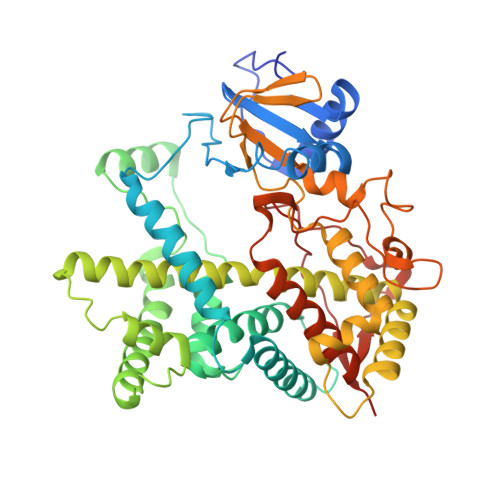
Structure of Cytochrome P450 2B4 with Bound Bifonazole


Experimental Data Snapshot

  • Method: X-RAY DIFFRACTION
  • Resolution: 2.30 Å
  • R-Value Free: 0.215 
  • R-Value Work: 0.196 
  • R-Value Observed: 0.200 

Starting Models: experimental
View more details

wwPDB Validation   3D Report Full Report


Ligand Structure Quality Assessment 


This is version 1.5 of the entry. See complete history


Literature

Structure of microsomal cytochrome P450 2B4 complexed with the antifungal drug bifonazole: insight into P450 conformational plasticity and membrane interaction.

Zhao, Y.White, M.A.Muralidhara, B.K.Sun, L.Halpert, J.R.Stout, C.D.

(2006) J Biol Chem 281: 5973-5981

  • DOI: https://doi.org/10.1074/jbc.M511464200
  • Primary Citation of Related Structures:  
    2BDM

  • PubMed Abstract: 

    To better understand ligand-induced structural transitions in cytochrome P450 2B4, protein-ligand interactions were investigated using a bulky inhibitor. Bifonazole, a broad spectrum antifungal agent, inhibits monooxygenase activity and induces a type II binding spectrum in 2B4dH(H226Y), a modified enzyme previously crystallized in the presence of 4-(4-chlorophenyl)imidazole (CPI). Isothermal titration calorimetry and tryptophan fluorescence quenching indicate no significant burial of protein apolar surface nor altered accessibility of Trp-121 upon bifonazole binding, in contrast to recent results with CPI. A 2.3 A crystal structure of 2B4-bifonazole reveals a novel open conformation with ligand bound in the active site, which is significantly different from either the U-shaped cleft of ligand-free 2B4 or the small active site pocket of 2B4-CPI. The O-shaped active site cleft of 2B4-bifonazole is widely open in the middle but narrow at the top. A bifonazole molecule occupies the bottom of the active site cleft, where helix I is bent approximately 15 degrees to accommodate the bulky ligand. The structure also defines unanticipated interactions between helix C residues and bifonazole, suggesting an important role of helix C in azole recognition by mammalian P450s. Comparison of the ligand-free 2B4 structure, the 2B4-CPI structure, and the 2B4-bifonazole structure identifies structurally plastic regions that undergo correlated conformational changes in response to ligand binding. The most plastic regions are putative membrane-binding motifs involved in substrate access or substrate binding. The results allow us to model the membrane-associated state of P450 and provide insight into how lipophilic substrates access the buried active site.


  • Organizational Affiliation

    Department of Pharmacology and Toxicology, University of Texas Medical Branch, Galveston, TX 77555, USA. [email protected]


Macromolecules
Find similar proteins by:  (by identity cutoff)  |  3D Structure
Entity ID: 1
MoleculeChains Sequence LengthOrganismDetailsImage
Cytochrome P450 2B4476Oryctolagus cuniculusMutation(s): 9 
Gene Names: CYP2B4
EC: 1.14.14.1
Membrane Entity: Yes 
UniProt
Find proteins for P00178 (Oryctolagus cuniculus)
Explore P00178 
Go to UniProtKB:  P00178
Entity Groups  
Sequence Clusters30% Identity50% Identity70% Identity90% Identity95% Identity100% Identity
UniProt GroupP00178
Sequence Annotations
Expand
  • Reference Sequence
Small Molecules
Ligands 3 Unique
IDChains Name / Formula / InChI Key2D Diagram3D Interactions
HEM
Query on HEM

Download Ideal Coordinates CCD File 
B [auth A]PROTOPORPHYRIN IX CONTAINING FE
C34 H32 Fe N4 O4
KABFMIBPWCXCRK-RGGAHWMASA-L
CM5
Query on CM5

Download Ideal Coordinates CCD File 
F [auth A],
G [auth A],
H [auth A],
I [auth A]
5-CYCLOHEXYL-1-PENTYL-BETA-D-MALTOSIDE
C23 H42 O11
RVTGFZGNOSKUDA-ZNGNCRBCSA-N
TMI
Query on TMI

Download Ideal Coordinates CCD File 
C [auth A],
D [auth A],
E [auth A]
1-[PHENYL-(4-PHENYLPHENYL)-METHYL]IMIDAZOLE
C22 H18 N2
OCAPBUJLXMYKEJ-QFIPXVFZSA-N
Experimental Data & Validation

Experimental Data

  • Method: X-RAY DIFFRACTION
  • Resolution: 2.30 Å
  • R-Value Free: 0.215 
  • R-Value Work: 0.196 
  • R-Value Observed: 0.200 
  • Space Group: P 61 2 2
Unit Cell:
Length ( Å )Angle ( ˚ )
a = 203.21α = 90
b = 203.21β = 90
c = 103.45γ = 120
Software Package:
Software NamePurpose
SCALAdata scaling
CNSrefinement
PDB_EXTRACTdata extraction
CCP4data scaling

Structure Validation

View Full Validation Report


Ligand Structure Quality Assessment 


Entry History 

Deposition Data

Revision History  (Full details and data files)

  • Version 1.0: 2005-12-27
    Type: Initial release
  • Version 1.1: 2008-05-01
    Changes: Version format compliance
  • Version 1.2: 2011-07-13
    Changes: Derived calculations, Version format compliance
  • Version 1.3: 2017-10-18
    Changes: Refinement description
  • Version 1.4: 2021-10-20
    Changes: Database references, Derived calculations
  • Version 1.5: 2023-08-23
    Changes: Data collection, Refinement description