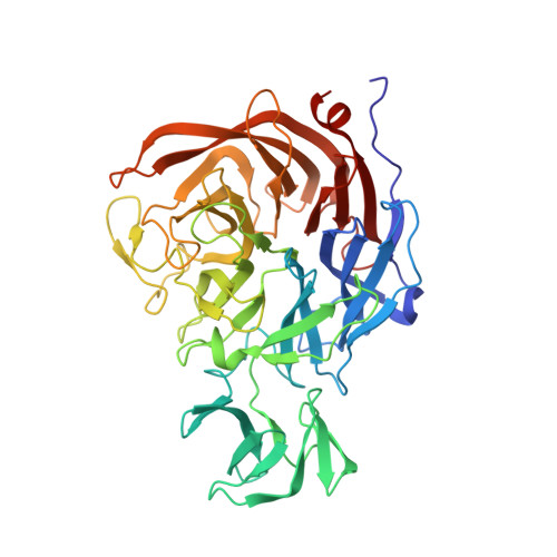
2BF6

Atomic Resolution Structure of the bacterial sialidase NanI from Clostridium perfringens in complex with alpha-Sialic Acid (Neu5Ac).


Experimental Data Snapshot

  • Method: X-RAY DIFFRACTION
  • Resolution: 0.97 Å
  • R-Value Free: 0.133 
  • R-Value Observed: 0.113 

Starting Model: experimental
View more details

wwPDB Validation   3D Report Full Report


Ligand Structure Quality Assessment 


This is version 1.6 of the entry. See complete history


Literature

The Structure of Clostridium Perfringens Nani Sialidase and its Catalytic Intermediates.

Newstead, S.L.Potter, J.A.Wilson, J.C.Xu, G.Chien, C.H.Watts, A.G.Withers, S.G.Taylor, G.L.

(2008) J Biol Chem 283: 9080

  • DOI: https://doi.org/10.1074/jbc.M710247200
  • Primary Citation of Related Structures:  
    2BF6, 2VK5, 2VK6, 2VK7

  • PubMed Abstract: 

    Clostridium perfringens is a Gram-positive bacterium responsible for bacteremia, gas gangrene, and occasionally food poisoning. Its genome encodes three sialidases, nanH, nanI, and nanJ, that are involved in the removal of sialic acids from a variety of glycoconjugates and that play a role in bacterial nutrition and pathogenesis. Recent studies on trypanosomal (trans-) sialidases have suggested that catalysis in all sialidases may proceed via a covalent intermediate similar to that of other retaining glycosidases. Here we provide further evidence to support this suggestion by reporting the 0.97A resolution atomic structure of the catalytic domain of the C. perfringens NanI sialidase, and complexes with its substrate sialic acid (N-acetylneuramic acid) also to 0.97A resolution, with a transition-state analogue (2-deoxy-2,3-dehydro-N-acetylneuraminic acid) to 1.5A resolution, and with a covalent intermediate formed using a fluorinated sialic acid analogue to 1.2A resolution. Together, these structures provide high resolution snapshots along the catalytic pathway. The crystal structures suggested that NanI is able to hydrate 2-deoxy-2,3-dehydro-N-acetylneuraminic acid to N-acetylneuramic acid. This was confirmed by NMR, and a mechanism for this activity is suggested.


  • Organizational Affiliation

    Centre for Biomolecular Sciences, University of St. Andrews, St. Andrews, Fife, United Kingdom.


Macromolecules
Find similar proteins by:  (by identity cutoff)  |  3D Structure
Entity ID: 1
MoleculeChains Sequence LengthOrganismDetailsImage
EXO-ALPHA-SIALIDASE449Clostridium perfringensMutation(s): 0 
EC: 3.2.1.18
UniProt
Find proteins for Q59310 (Clostridium perfringens)
Explore Q59310 
Go to UniProtKB:  Q59310
Entity Groups  
Sequence Clusters30% Identity50% Identity70% Identity90% Identity95% Identity100% Identity
UniProt GroupQ59310
Sequence Annotations
Expand
  • Reference Sequence
Experimental Data & Validation

Experimental Data

  • Method: X-RAY DIFFRACTION
  • Resolution: 0.97 Å
  • R-Value Free: 0.133 
  • R-Value Observed: 0.113 
  • Space Group: P 21 21 21
Unit Cell:
Length ( Å )Angle ( ˚ )
a = 96.981α = 90
b = 69.413β = 90
c = 72.694γ = 90
Software Package:
Software NamePurpose
SHELXL-97refinement
MOSFLMdata reduction
SCALAdata scaling
CNSphasing

Structure Validation

View Full Validation Report


Ligand Structure Quality Assessment 


Entry History 

Deposition Data

Revision History  (Full details and data files)

  • Version 1.0: 2006-03-09
    Type: Initial release
  • Version 1.1: 2011-05-08
    Changes: Version format compliance
  • Version 1.2: 2011-07-13
    Changes: Version format compliance
  • Version 1.3: 2018-03-07
    Changes: Advisory, Source and taxonomy
  • Version 1.4: 2019-05-22
    Changes: Data collection, Refinement description
  • Version 1.5: 2020-07-29
    Type: Remediation
    Reason: Carbohydrate remediation
    Changes: Data collection, Derived calculations, Other, Structure summary
  • Version 1.6: 2023-12-13
    Changes: Advisory, Data collection, Database references, Refinement description, Structure summary