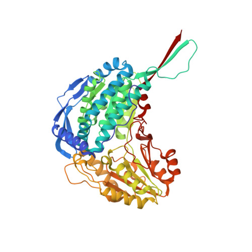
2BHQ

Crystal Analysis of 1-Pyrroline-5-Carboxylate Dehydrogenase from Thermus with bound product glutamate.


Experimental Data Snapshot

  • Method: X-RAY DIFFRACTION
  • Resolution: 1.40 Å
  • R-Value Free: 0.186 
  • R-Value Work: 0.170 
  • R-Value Observed: 0.170 

Starting Model: experimental
View more details

wwPDB Validation   3D Report Full Report


This is version 1.3 of the entry. See complete history


Literature

Crystal Structure of Thermus Thermophilus Delta(1)- Pyrroline-5-Carboxylate Dehydrogenase.

Inagaki, E.Ohshima, N.Takahashi, H.Kuroishi, C.Yokoyama, S.Tahirov, T.H.

(2006) J Mol Biol 362: 490

  • DOI: https://doi.org/10.1016/j.jmb.2006.07.048
  • Primary Citation of Related Structures:  
    1UZB, 2BHP, 2BHQ, 2BJA, 2BJK, 2IY6

  • PubMed Abstract: 

    Delta(1)-pyrroline-5-carboxylate dehydrogenase (P5CDh) plays an important role in the metabolic pathway from proline to glutamate. It irreversibly catalyzes the oxidation of glutamate-gamma-semialdehyde, the product of the non-enzymatic hydrolysis of Delta(1)-pyrroline-5-carboxylate, into glutamate with the reduction of NAD(+) into NADH. We have confirmed the P5CDh activity of the Thermus thermophilus protein TT0033 (TtP5CDh), and determined the crystal structure of the enzyme in the ligand-free form at 1.4 A resolution. To investigate the structural basis of TtP5CDh function, the TtP5CDh structures with NAD(+), with NADH, and with its product glutamate were determined at 1.8 A, 1.9 A, and 1.4 A resolution, respectively. The solved structures suggest an overall view of the P5CDh catalytic mechanism and provide insights into the P5CDh deficiencies in the case of the human type II hyperprolinemia.


  • Organizational Affiliation

    RIKEN SPring-8 Center, Harima Institute, 1-1-1 Kouto, Sayo, Hyogo 679-5148, Japan.


Macromolecules
Find similar proteins by:  (by identity cutoff)  |  3D Structure
Entity ID: 1
MoleculeChains Sequence LengthOrganismDetailsImage
1-PYRROLINE-5-CARBOXYLATE DEHYDROGENASE
A, B
516Thermus thermophilus HB8Mutation(s): 0 
EC: 1.5.1.12 (PDB Primary Data), 1.2.1.88 (UniProt)
UniProt
Find proteins for Q5SI02 (Thermus thermophilus (strain ATCC 27634 / DSM 579 / HB8))
Explore Q5SI02 
Go to UniProtKB:  Q5SI02
Entity Groups  
Sequence Clusters30% Identity50% Identity70% Identity90% Identity95% Identity100% Identity
UniProt GroupQ5SI02
Sequence Annotations
Expand
  • Reference Sequence
Small Molecules
Ligands 4 Unique
IDChains Name / Formula / InChI Key2D Diagram3D Interactions
GLU
Query on GLU

Download Ideal Coordinates CCD File 
C [auth A],
K [auth B]
GLUTAMIC ACID
C5 H9 N O4
WHUUTDBJXJRKMK-VKHMYHEASA-N
MPD
Query on MPD

Download Ideal Coordinates CCD File 
F [auth A],
G [auth A],
I [auth A],
N [auth B]
(4S)-2-METHYL-2,4-PENTANEDIOL
C6 H14 O2
SVTBMSDMJJWYQN-YFKPBYRVSA-N
MRD
Query on MRD

Download Ideal Coordinates CCD File 
D [auth A],
E [auth A],
H [auth A],
L [auth B],
O [auth B]
(4R)-2-METHYLPENTANE-2,4-DIOL
C6 H14 O2
SVTBMSDMJJWYQN-RXMQYKEDSA-N
ACT
Query on ACT

Download Ideal Coordinates CCD File 
J [auth A],
M [auth B]
ACETATE ION
C2 H3 O2
QTBSBXVTEAMEQO-UHFFFAOYSA-M
Experimental Data & Validation

Experimental Data

  • Method: X-RAY DIFFRACTION
  • Resolution: 1.40 Å
  • R-Value Free: 0.186 
  • R-Value Work: 0.170 
  • R-Value Observed: 0.170 
  • Space Group: H 3
Unit Cell:
Length ( Å )Angle ( ˚ )
a = 102.618α = 90
b = 102.618β = 90
c = 279.074γ = 120
Software Package:
Software NamePurpose
CNSrefinement
HKL-2000data reduction
SCALEPACKdata scaling
CNSphasing

Structure Validation

View Full Validation Report


Entry History 

Deposition Data

Revision History  (Full details and data files)

  • Version 1.0: 2006-03-09
    Type: Initial release
  • Version 1.1: 2011-05-08
    Changes: Version format compliance
  • Version 1.2: 2011-07-13
    Changes: Version format compliance
  • Version 1.3: 2023-12-13
    Changes: Data collection, Database references, Other, Refinement description