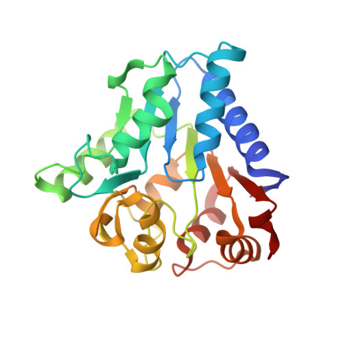
2BKV

Structure and kinetics of a monomeric glucosamine-6-phosphate deaminase: missing link of the NagB superfamily


Experimental Data Snapshot

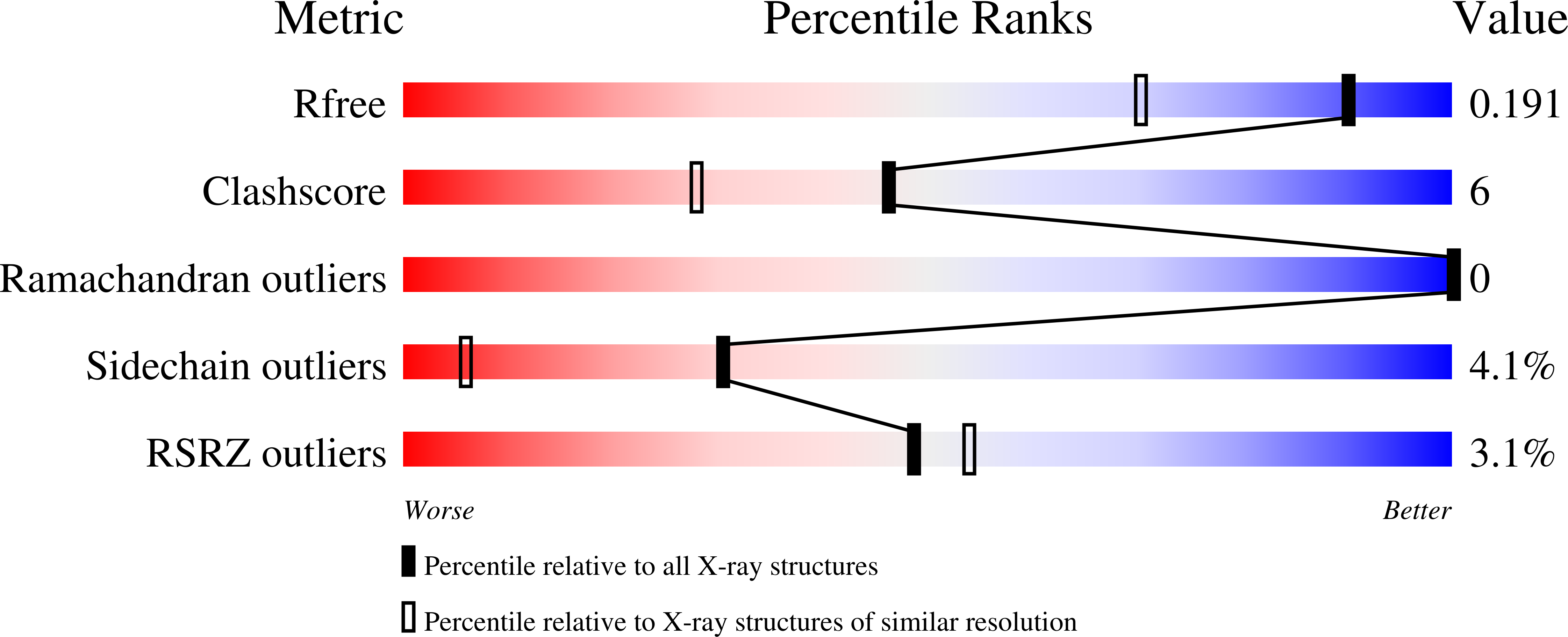
  • Method: X-RAY DIFFRACTION
  • Resolution: 1.50 Å
  • R-Value Free: 0.211 
  • R-Value Work: 0.165 
  • R-Value Observed: 0.167 

Starting Model: experimental
View more details

wwPDB Validation   3D Report Full Report


Ligand Structure Quality Assessment 


This is version 1.4 of the entry. See complete history


Literature

Structure and Kinetics of a Monomeric Glucosamine 6-Phosphate Deaminase: Missing Link of the Nagb Superfamily

Vincent, F.Davies, G.J.Brannigan, J.A.

(2005) J Biol Chem 280: 19649

  • DOI: https://doi.org/10.1074/jbc.M502131200
  • Primary Citation of Related Structures:  
    2BKV, 2BKX

  • PubMed Abstract: 

    Glucosamine 6-phosphate is converted to fructose 6-phosphate and ammonia by the action of the enzyme glucosamine 6-phosphate deaminase, NagB. This reaction is the final step in the specific GlcNAc utilization pathway and thus decides the metabolic fate of GlcNAc. Sequence analyses suggest that the NagB "superfamily" consists of three main clusters: multimeric and allosterically regulated glucosamine-6-phosphate deaminases (exemplified by Escherichia coli NagB), phosphogluconolactonases, and monomeric hexosamine-6-phosphate deaminases. Here we present the three-dimensional structure and kinetics of the first member of this latter group, the glucosamine-6-phosphate deaminase, NagB, from Bacillus subtilis. The structures were determined in ligand-complexed forms at resolutions around 1.4 Angstroms. BsuNagB is monomeric in solution and as a consequence is active (k(cat) 28 s(-1), K(m(app)) 0.13 mM) without the need for allosteric activators. A decrease in activity at high substrate concentrations may reflect substrate inhibition (with K(i) of approximately 4 mM). The structure completes the NagB superfamily structural landscape and thus allows further interrogation of genomic data in terms of the regulation of NagB and the metabolic fate(s) of glucosamine 6-phosphate.


  • Organizational Affiliation

    Structural Biology Laboratory, Department of Chemistry, University of York, Heslington, UK.


Macromolecules
Find similar proteins by:  (by identity cutoff)  |  3D Structure
Entity ID: 1
MoleculeChains Sequence LengthOrganismDetailsImage
GLUCOSAMINE-6-PHOSPHATE DEAMINASE
A, B
242Bacillus subtilisMutation(s): 0 
EC: 3.5.99.6
UniProt
Find proteins for O35000 (Bacillus subtilis (strain 168))
Explore O35000 
Go to UniProtKB:  O35000
Entity Groups  
Sequence Clusters30% Identity50% Identity70% Identity90% Identity95% Identity100% Identity
UniProt GroupO35000
Sequence Annotations
Expand
  • Reference Sequence
Experimental Data & Validation

Experimental Data

  • Method: X-RAY DIFFRACTION
  • Resolution: 1.50 Å
  • R-Value Free: 0.211 
  • R-Value Work: 0.165 
  • R-Value Observed: 0.167 
  • Space Group: P 1 21 1
Unit Cell:
Length ( Å )Angle ( ˚ )
a = 63.009α = 90
b = 48.023β = 91.04
c = 71.761γ = 90
Software Package:
Software NamePurpose
REFMACrefinement
HKL-2000data reduction
SCALEPACKdata scaling
AMoREphasing

Structure Validation

View Full Validation Report


Ligand Structure Quality Assessment 


Entry History 

Deposition Data

Revision History  (Full details and data files)

  • Version 1.0: 2005-03-09
    Type: Initial release
  • Version 1.1: 2011-05-08
    Changes: Version format compliance
  • Version 1.2: 2011-07-13
    Changes: Version format compliance
  • Version 1.3: 2023-12-13
    Changes: Data collection, Database references, Other, Refinement description
  • Version 1.4: 2024-05-01
    Changes: Advisory