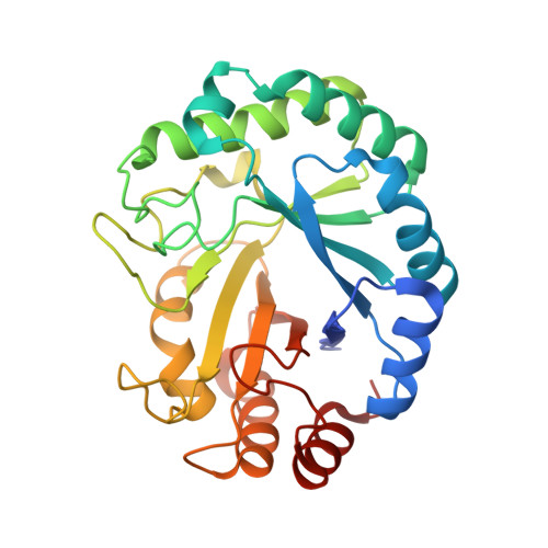
2BVD

HOW FAMILY 26 GLYCOSIDE HYDROLASES ORCHESTRATE CATALYSIS ON DIFFERENT POLYSACCHARIDES. STRUCTURE AND ACTIVITY OF A CLOSTRIDIUM THERMOCELLUM LICHENASE, CtLIC26A


Experimental Data Snapshot

  • Method: X-RAY DIFFRACTION
  • Resolution: 1.60 Å
  • R-Value Free: 0.186 
  • R-Value Work: 0.154 
  • R-Value Observed: 0.156 

Starting Model: experimental
View more details

wwPDB Validation   3D Report Full Report


Ligand Structure Quality Assessment 


This is version 2.1 of the entry. See complete history


Literature

How Family 26 Glycoside Hydrolases Orchestrate Catalysis on Different Polysaccharides: Structure and Activity of a Clostridium Thermocellum Lichenase, Ctlic26A.

Taylor, E.J.Goyal, A.Guerreiro, C.I.P.D.Prates, J.A.M.Money, V.A.Ferry, N.Morland, C.Planas, A.Macdonald, J.A.Stick, R.V.Gilbert, H.J.Fontes, C.M.G.A.Davies, G.J.

(2005) J Biol Chem 280: 32761

  • DOI: https://doi.org/10.1074/jbc.M506580200
  • Primary Citation of Related Structures:  
    2BV9, 2BVD

  • PubMed Abstract: 

    One of the most intriguing features of the 90 glycoside hydrolase families (GHs) is the range of specificities displayed by different members of the same family, whereas the catalytic apparatus and mechanism are often invariant. Family GH26 predominantly comprises beta-1,4 mannanases; however, a bifunctional Clostridium thermocellum GH26 member (hereafter CtLic26A) displays a markedly different specificity. We show that CtLic26A is a lichenase, specific for mixed (Glcbeta1,4Glcbeta1,4Glcbeta1,3)n oligo- and polysaccharides, and displays no activity on manno-configured substrates or beta-1,4-linked homopolymers of glucose or xylose. The three-dimensional structure of the native form of CtLic26A has been solved at 1.50-A resolution, revealing a characteristic (beta/alpha)8 barrel with Glu-109 and Glu-222 acting as the catalytic acid/base and nucleophile in a double-displacement mechanism. The complex with the competitive inhibitor, Glc-beta-1,3-isofagomine (Ki 1 microm), at 1.60 A sheds light on substrate recognition in the -2 and -1 subsites and illuminates why the enzyme is specific for lichenan-based substrates. Hydrolysis of beta-mannosides by GH26 members is thought to proceed through transition states in the B2,5 (boat) conformation in which structural distinction of glucosides versus mannosides reflects not the configuration at C2 but the recognition of the pseudoaxial O3 of the B2,5 conformation. We suggest a different conformational itinerary for the GH26 enzymes active on gluco-configured substrates.


  • Organizational Affiliation

    York Structural Biology Laboratory, Department of Chemistry, University of York, York, YO10 5YW, United Kingdom.


Macromolecules
Find similar proteins by:  (by identity cutoff)  |  3D Structure
Entity ID: 1
MoleculeChains Sequence LengthOrganismDetailsImage
ENDOGLUCANASE H283Acetivibrio thermocellusMutation(s): 0 
EC: 3.2.1.4
UniProt
Find proteins for P16218 (Acetivibrio thermocellus (strain ATCC 27405 / DSM 1237 / JCM 9322 / NBRC 103400 / NCIMB 10682 / NRRL B-4536 / VPI 7372))
Explore P16218 
Go to UniProtKB:  P16218
Entity Groups  
Sequence Clusters30% Identity50% Identity70% Identity90% Identity95% Identity100% Identity
UniProt GroupP16218
Sequence Annotations
Expand
  • Reference Sequence
Small Molecules
Ligands 1 Unique
IDChains Name / Formula / InChI Key2D Diagram3D Interactions
ISX
Query on ISX

Download Ideal Coordinates CCD File 
B [auth A](3R,4R,5R)-4-hydroxy-5-(hydroxymethyl)piperidin-3-yl beta-D-glucopyranoside
C12 H23 N O8
BEXVSXHKIARRKH-FYKVHUBJSA-N
Binding Affinity Annotations 
IDSourceBinding Affinity
ISX PDBBind:  2BVD Ki: 1000 (nM) from 1 assay(s)
Experimental Data & Validation

Experimental Data

  • Method: X-RAY DIFFRACTION
  • Resolution: 1.60 Å
  • R-Value Free: 0.186 
  • R-Value Work: 0.154 
  • R-Value Observed: 0.156 
  • Space Group: P 21 21 21
Unit Cell:
Length ( Å )Angle ( ˚ )
a = 49.272α = 90
b = 63.012β = 90
c = 78.182γ = 90
Software Package:
Software NamePurpose
REFMACrefinement
MOSFLMdata reduction
SCALAdata scaling
AMoREphasing

Structure Validation

View Full Validation Report


Ligand Structure Quality Assessment 


Entry History 

Revision History  (Full details and data files)

  • Version 1.0: 2005-06-30
    Type: Initial release
  • Version 1.1: 2011-05-08
    Changes: Version format compliance
  • Version 1.2: 2011-07-13
    Changes: Version format compliance
  • Version 2.0: 2020-07-29
    Type: Remediation
    Reason: Carbohydrate remediation
    Changes: Atomic model, Derived calculations, Other, Structure summary
  • Version 2.1: 2023-12-13
    Changes: Data collection, Database references, Refinement description, Structure summary