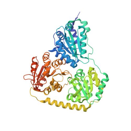
2C31

CRYSTAL STRUCTURE OF OXALYL-COA DECARBOXYLASE IN COMPLEX WITH THE COFACTOR DERIVATIVE THIAMIN-2-THIAZOLONE DIPHOSPHATE AND ADENOSINE DIPHOSPHATE


Experimental Data Snapshot

  • Method: X-RAY DIFFRACTION
  • Resolution: 1.73 Å
  • R-Value Free: 0.175 
  • R-Value Work: 0.150 
  • R-Value Observed: 0.150 

Starting Model: experimental
View more details

wwPDB Validation   3D Report Full Report


Ligand Structure Quality Assessment 


This is version 1.3 of the entry. See complete history


Literature

Structural Basis for Activation of the Thiamin Diphosphate-Dependent Enzyme Oxalyl-Coa Decarboxylase by Adenosine Diphosphate.

Berthold, C.L.Moussatche, P.Richards, N.G.J.Lindqvist, Y.

(2005) J Biol Chem 280: 41645

  • DOI: https://doi.org/10.1074/jbc.M509921200
  • Primary Citation of Related Structures:  
    2C31

  • PubMed Abstract: 

    Oxalyl-coenzyme A decarboxylase is a thiamin diphosphate-dependent enzyme that plays an important role in the catabolism of the highly toxic compound oxalate. We have determined the crystal structure of the enzyme from Oxalobacter formigenes from a hemihedrally twinned crystal to 1.73 A resolution and characterized the steady-state kinetic behavior of the decarboxylase. The monomer of the tetrameric enzyme consists of three alpha/beta-type domains, commonly seen in this class of enzymes, and the thiamin diphosphate-binding site is located at the expected subunit-subunit interface between two of the domains with the cofactor bound in the conserved V-conformation. Although oxalyl-CoA decarboxylase is structurally homologous to acetohydroxyacid synthase, a molecule of ADP is bound in a region that is cognate to the FAD-binding site observed in acetohydroxyacid synthase and presumably fulfils a similar role in stabilizing the protein structure. This difference between the two enzymes may have physiological importance since oxalyl-CoA decarboxylation is an essential step in ATP generation in O. formigenes, and the decarboxylase activity is stimulated by exogenous ADP. Despite the significant degree of structural conservation between the two homologous enzymes and the similarity in catalytic mechanism to other thiamin diphosphate-dependent enzymes, the active site residues of oxalyl-CoA decarboxylase are unique. A suggestion for the reaction mechanism of the enzyme is presented.


  • Organizational Affiliation

    Molecular Structural Biology, Department of Medical Biochemistry and Biophysics, Karolinska Institutet, S-17177 Stockholm, Sweden.


Macromolecules
Find similar proteins by:  (by identity cutoff)  |  3D Structure
Entity ID: 1
MoleculeChains Sequence LengthOrganismDetailsImage
OXALYL-COA DECARBOXYLASE
A, B
568Oxalobacter formigenesMutation(s): 0 
EC: 4.1.1.8
UniProt
Find proteins for P40149 (Oxalobacter formigenes)
Explore P40149 
Go to UniProtKB:  P40149
Entity Groups  
Sequence Clusters30% Identity50% Identity70% Identity90% Identity95% Identity100% Identity
UniProt GroupP40149
Sequence Annotations
Expand
  • Reference Sequence
Small Molecules
Ligands 3 Unique
IDChains Name / Formula / InChI Key2D Diagram3D Interactions
TZD
Query on TZD

Download Ideal Coordinates CCD File 
D [auth A],
G [auth B]
2-{3-[(4-AMINO-2-METHYLPYRIMIDIN-5-YL)METHYL]-4-METHYL-2-OXO-2,3-DIHYDRO-1,3-THIAZOL-5-YL}ETHYL TRIHYDROGEN DIPHOSPHATE
C12 H18 N4 O8 P2 S
ZGJUYGIRPQSCFA-UHFFFAOYSA-N
ADP
Query on ADP

Download Ideal Coordinates CCD File 
E [auth A],
H [auth B]
ADENOSINE-5'-DIPHOSPHATE
C10 H15 N5 O10 P2
XTWYTFMLZFPYCI-KQYNXXCUSA-N
MG
Query on MG

Download Ideal Coordinates CCD File 
C [auth A],
F [auth B]
MAGNESIUM ION
Mg
JLVVSXFLKOJNIY-UHFFFAOYSA-N
Experimental Data & Validation

Experimental Data

  • Method: X-RAY DIFFRACTION
  • Resolution: 1.73 Å
  • R-Value Free: 0.175 
  • R-Value Work: 0.150 
  • R-Value Observed: 0.150 
  • Space Group: P 31 2 1
Unit Cell:
Length ( Å )Angle ( ˚ )
a = 124.3α = 90
b = 124.3β = 90
c = 151.7γ = 120
Software Package:
Software NamePurpose
CNSrefinement
MOSFLMdata reduction
SCALAdata scaling
MOLREPphasing

Structure Validation

View Full Validation Report


Ligand Structure Quality Assessment 


Entry History 

Deposition Data

Revision History  (Full details and data files)

  • Version 1.0: 2005-10-07
    Type: Initial release
  • Version 1.1: 2011-05-07
    Changes: Version format compliance
  • Version 1.2: 2011-07-13
    Changes: Version format compliance
  • Version 1.3: 2023-12-13
    Changes: Data collection, Database references, Derived calculations, Other, Refinement description