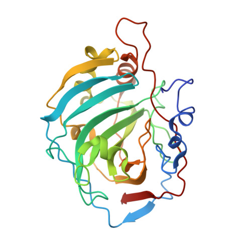
2CAB

STRUCTURE, REFINEMENT AND FUNCTION OF CARBONIC ANHYDRASE ISOZYMES. REFINEMENT OF HUMAN CARBONIC ANHYDRASE I


Experimental Data Snapshot

  • Method: X-RAY DIFFRACTION
  • Resolution: 2.00 Å
  • R-Value Work: 0.193 

wwPDB Validation   3D Report Full Report


This is version 1.4 of the entry. See complete history


Literature

Structure, refinement, and function of carbonic anhydrase isozymes: refinement of human carbonic anhydrase I

Kannan, K.K.Ramanadham, M.Jones, T.A.

(1984) Ann N Y Acad Sci 429: 49-60

  • DOI: https://doi.org/10.1111/j.1749-6632.1984.tb12314.x
  • Primary Citation of Related Structures:  
    2CAB

  • PubMed Abstract: 

    The structure of human erythrocyte carbonic anhydrase I has been refined to a final R value of 19% to 2-A resolution by a combination of least squares refinement and model fitting in a three-dimensional graphics display. About 300 solvent atoms have been located bound to the protein molecule. An interesting hydrogen bond network involving Zn2+, the liganded solvent, side chain groups of Thr-199, Glu-106, Thr-7, and His-64 through two solvent molecules have been found that may be important for the catalytic mechanism of the carbonic anhydrase.


Macromolecules
Find similar proteins by:  (by identity cutoff)  |  3D Structure
Entity ID: 1
MoleculeChains Sequence LengthOrganismDetailsImage
CARBONIC ANHYDRASE FORM B260Homo sapiensMutation(s): 0 
EC: 4.2.1.1 (PDB Primary Data), 4.2.1.69 (UniProt)
UniProt & NIH Common Fund Data Resources
Find proteins for P00915 (Homo sapiens)
Explore P00915 
Go to UniProtKB:  P00915
PHAROS:  P00915
GTEx:  ENSG00000133742 
Entity Groups  
Sequence Clusters30% Identity50% Identity70% Identity90% Identity95% Identity100% Identity
UniProt GroupP00915
Sequence Annotations
Expand
  • Reference Sequence
Small Molecules
Ligands 1 Unique
IDChains Name / Formula / InChI Key2D Diagram3D Interactions
ZN
Query on ZN

Download Ideal Coordinates CCD File 
B [auth A]ZINC ION
Zn
PTFCDOFLOPIGGS-UHFFFAOYSA-N
Experimental Data & Validation

Experimental Data

  • Method: X-RAY DIFFRACTION
  • Resolution: 2.00 Å
  • R-Value Work: 0.193 
  • Space Group: P 21 21 21
Unit Cell:
Length ( Å )Angle ( ˚ )
a = 81.5α = 90
b = 73.6β = 90
c = 37.1γ = 90
Software Package:
Software NamePurpose
CORELSrefinement

Structure Validation

View Full Validation Report


Entry History 

Deposition Data

Revision History  (Full details and data files)

  • Version 1.0: 1984-02-02
    Type: Initial release
  • Version 1.1: 2008-03-24
    Changes: Version format compliance
  • Version 1.2: 2011-07-13
    Changes: Version format compliance
  • Version 1.3: 2017-11-29
    Changes: Derived calculations, Other
  • Version 1.4: 2024-02-14
    Changes: Data collection, Database references, Derived calculations