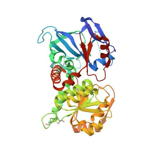
2CDB

Sulfolobus solfataricus Glucose Dehydrogenase 1 in complex with NADP and glucose


Experimental Data Snapshot

  • Method: X-RAY DIFFRACTION
  • Resolution: 1.60 Å
  • R-Value Free: 0.218 
  • R-Value Work: 0.190 
  • R-Value Observed: 0.192 

Starting Model: experimental
View more details

wwPDB Validation   3D Report Full Report


Ligand Structure Quality Assessment 


This is version 1.4 of the entry. See complete history


Literature

The Structural Basis of Substrate Promiscuity in Glucose Dehydrogenase from the Hyperthermophilic Archaeon Sulfolobus Solfataricus.

Milburn, C.C.Lamble, H.J.Theodossis, A.Bull, S.D.Hough, D.W.Danson, M.J.Taylor, G.L.

(2006) J Biol Chem 281: 14796

  • DOI: https://doi.org/10.1074/jbc.M601334200
  • Primary Citation of Related Structures:  
    2CD9, 2CDA, 2CDB, 2CDC

  • PubMed Abstract: 

    The hyperthermophilic archaeon Sulfolobus solfataricus grows optimally above 80 degrees C and utilizes an unusual, promiscuous, non-phosphorylative Entner-Doudoroff pathway to metabolize both glucose and galactose. The first enzyme in this pathway, glucose dehydrogenase, catalyzes the oxidation of glucose to gluconate, but has been shown to have activity with a broad range of sugar substrates, including glucose, galactose, xylose, and L-arabinose, with a requirement for the glucose stereo configuration at the C2 and C3 positions. Here we report the crystal structure of the apo form of glucose dehydrogenase to a resolution of 1.8 A and a complex with its required cofactor, NADP+, to a resolution of 2.3 A. A T41A mutation was engineered to enable the trapping of substrate in the crystal. Complexes of the enzyme with D-glucose and D-xylose are presented to resolutions of 1.6 and 1.5 A, respectively, that provide evidence of selectivity for the beta-anomeric, pyranose form of the substrate, and indicate that this is the productive substrate form. The nature of the promiscuity of glucose dehydrogenase is also elucidated, and a physiological role for this enzyme in xylose metabolism is suggested. Finally, the structure suggests that the mechanism of sugar oxidation by this enzyme may be similar to that described for human sorbitol dehydrogenase.


  • Organizational Affiliation

    Centre for Biomolecular Sciences, University of St. Andrews, North Haugh, St. Andrews, Fife KY16 9ST, Scotland, UK.


Macromolecules
Find similar proteins by:  (by identity cutoff)  |  3D Structure
Entity ID: 1
MoleculeChains Sequence LengthOrganismDetailsImage
GLUCOSE 1-DEHYDROGENASE (DHG-1)
A, B, C, D
366Saccharolobus solfataricusMutation(s): 1 
EC: 1.1.1.47 (PDB Primary Data), 1.1.1.359 (UniProt), 1.1.1.48 (UniProt), 1.1.1.120 (UniProt)
UniProt
Find proteins for O93715 (Saccharolobus solfataricus)
Explore O93715 
Go to UniProtKB:  O93715
Entity Groups  
Sequence Clusters30% Identity50% Identity70% Identity90% Identity95% Identity100% Identity
UniProt GroupO93715
Sequence Annotations
Expand
  • Reference Sequence
Small Molecules
Ligands 4 Unique
IDChains Name / Formula / InChI Key2D Diagram3D Interactions
NAP
Query on NAP

Download Ideal Coordinates CCD File 
G [auth A],
L [auth B],
Q [auth C],
V [auth D]
NADP NICOTINAMIDE-ADENINE-DINUCLEOTIDE PHOSPHATE
C21 H28 N7 O17 P3
XJLXINKUBYWONI-NNYOXOHSSA-N
BGC
Query on BGC

Download Ideal Coordinates CCD File 
F [auth A],
K [auth B],
P [auth C],
U [auth D]
beta-D-glucopyranose
C6 H12 O6
WQZGKKKJIJFFOK-VFUOTHLCSA-N
ZN
Query on ZN

Download Ideal Coordinates CCD File 
H [auth A]
I [auth A]
M [auth B]
N [auth B]
R [auth C]
H [auth A],
I [auth A],
M [auth B],
N [auth B],
R [auth C],
S [auth C],
W [auth D],
X [auth D]
ZINC ION
Zn
PTFCDOFLOPIGGS-UHFFFAOYSA-N
EDO
Query on EDO

Download Ideal Coordinates CCD File 
E [auth A],
J [auth B],
O [auth C],
T [auth D]
1,2-ETHANEDIOL
C2 H6 O2
LYCAIKOWRPUZTN-UHFFFAOYSA-N
Experimental Data & Validation

Experimental Data

  • Method: X-RAY DIFFRACTION
  • Resolution: 1.60 Å
  • R-Value Free: 0.218 
  • R-Value Work: 0.190 
  • R-Value Observed: 0.192 
  • Space Group: P 1 21 1
Unit Cell:
Length ( Å )Angle ( ˚ )
a = 68.672α = 90
b = 91.394β = 90.03
c = 138.433γ = 90
Software Package:
Software NamePurpose
REFMACrefinement
MOSFLMdata reduction
SCALAdata scaling
AMoREphasing

Structure Validation

View Full Validation Report


Ligand Structure Quality Assessment 


Entry History 

Deposition Data

Revision History  (Full details and data files)

  • Version 1.0: 2006-03-22
    Type: Initial release
  • Version 1.1: 2011-07-13
    Changes: Advisory, Refinement description, Version format compliance
  • Version 1.2: 2020-07-29
    Type: Remediation
    Reason: Carbohydrate remediation
    Changes: Advisory, Data collection, Derived calculations, Other, Structure summary
  • Version 1.3: 2023-12-13
    Changes: Data collection, Database references, Refinement description, Structure summary
  • Version 1.4: 2024-11-13
    Changes: Structure summary