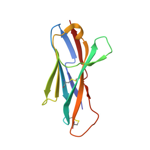
2COQ

Structure of new antigen receptor variable domain from sharks


Experimental Data Snapshot

  • Method: X-RAY DIFFRACTION
  • Resolution: 2.10 Å
  • R-Value Free: 0.280 
  • R-Value Work: 0.218 
  • R-Value Observed: 0.220 

Starting Model: experimental
View more details

wwPDB Validation   3D Report Full Report


This is version 1.4 of the entry. See complete history


Literature

Structure of a shark IgNAR antibody variable domain and modeling of an early-developmental isotype.

Streltsov, V.A.Carmichael, J.A.Nuttall, S.D.

(2005) Protein Sci 14: 2901-2909

  • DOI: https://doi.org/10.1110/ps.051709505
  • Primary Citation of Related Structures:  
    2COQ

  • PubMed Abstract: 

    The new antigen receptor (IgNAR) antibodies from sharks are disulphide bonded dimers of two protein chains, each containing one variable and five constant domains. Three types of IgNAR variable domains have been discovered, with Type 3 appearing early in shark development and being overtaken by the antigen-driven affinity-matured Type 1 and 2 response. Here, we have determined the first structure of a naturally occurring Type 2 IgNAR variable domain, and identified the disulphide bond that links and stabilizes the CDR1 and CDR3 loops. This disulphide bridge locks the CDR3 loop in an "upright" conformation in contrast to other shark antibody structures, where a more lateral configuration is observed. Further, we sought to model the Type 3 isotype based on the crystallographic structure reported here. This modeling indicates (1) that internal Type 3-specific residues combine to pack into a compact immunoglobulin core that supports the CDR loop regions, and (2) that despite apparent low-sequence variability, there is sufficient plasticity in the CDR3 loop to form a conformationally diverse antigen-binding surface.


  • Organizational Affiliation

    CSIRO Molecular and Health Technologies, 343 Royal Parade, Parkville, Victoria 3052, Australia. [email protected]


Macromolecules
Find similar proteins by:  (by identity cutoff)  |  3D Structure
Entity ID: 1
MoleculeChains Sequence LengthOrganismDetailsImage
new antigen receptor variable domain108Orectolobus maculatusMutation(s): 0 
UniProt
Find proteins for Q8JJ25 (Orectolobus maculatus)
Explore Q8JJ25 
Go to UniProtKB:  Q8JJ25
Entity Groups  
Sequence Clusters30% Identity50% Identity70% Identity90% Identity95% Identity100% Identity
UniProt GroupQ8JJ25
Sequence Annotations
Expand
  • Reference Sequence
Experimental Data & Validation

Experimental Data

  • Method: X-RAY DIFFRACTION
  • Resolution: 2.10 Å
  • R-Value Free: 0.280 
  • R-Value Work: 0.218 
  • R-Value Observed: 0.220 
  • Space Group: P 21 21 2
Unit Cell:
Length ( Å )Angle ( ˚ )
a = 38.268α = 90
b = 68.324β = 90
c = 39.511γ = 90
Software Package:
Software NamePurpose
DENZOdata reduction
SCALEPACKdata scaling
CCP4model building
REFMACrefinement
MOLREPphasing

Structure Validation

View Full Validation Report


Entry History 

Deposition Data

Revision History  (Full details and data files)

  • Version 1.0: 2005-10-25
    Type: Initial release
  • Version 1.1: 2008-04-30
    Changes: Version format compliance
  • Version 1.2: 2011-07-13
    Changes: Advisory, Version format compliance
  • Version 1.3: 2023-10-25
    Changes: Data collection, Database references, Refinement description
  • Version 1.4: 2024-10-23
    Changes: Structure summary