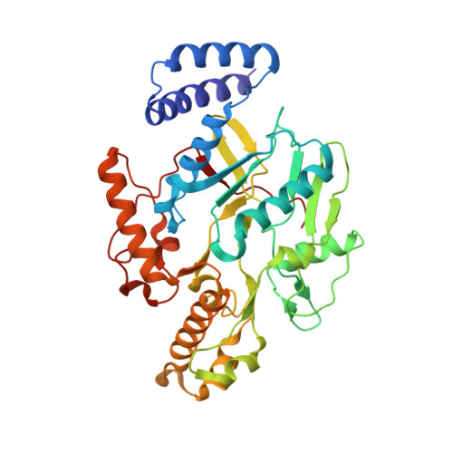
Nitrosyl-Heme Structures of Bacillus subtilis Nitric Oxide Synthase Have Implications for Understanding Substrate Oxidation.
Pant, K., Crane, B.R.(2006) Biochemistry 45: 2537-2544
- PubMed: 16489746
- DOI: https://doi.org/10.1021/bi0518848
- Primary Citation of Related Structures:
2FBZ, 2FC1, 2FC2 - PubMed Abstract:
The crystal structures of nitrosyl-heme complexes of a prokaryotic nitric oxide synthase (NOS) from Bacillus subtilis (bsNOS) reveal changes in active-site hydrogen bonding in the presence of the intermediate N(omega)-hydroxy-l-arginine (NOHA) compared to the substrate l-arginine (l-Arg). Correlating with a Val-to-Ile residue substitution in the bsNOS heme pocket, the Fe(II)-NO complex with both l-Arg and NOHA is more bent than the Fe(II)-NO, l-Arg complex of mammalian eNOS [Li, H., Raman, C. S., Martasek, P., Masters, B. S. S., and Poulos, T. L. (2001) Biochemistry 40, 5399-5406]. Structures of the Fe(III)-NO complex with NOHA show a nearly linear nitrosyl group, and in one subunit, partial nitrosation of bound NOHA. In the Fe(II)-NO complexes, the protonated NOHA N(omega) atom forms a short hydrogen bond with the heme-coordinated NO nitrogen, but active-site water molecules are out of hydrogen bonding range with the distal NO oxygen. In contrast, the l-Arg guanidinium interacts more weakly and equally with both NO atoms, and an active-site water molecule hydrogen bonds to the distal NO oxygen. This difference in hydrogen bonding to the nitrosyl group by the two substrates indicates that interactions provided by NOHA may preferentially stabilize an electrophilic peroxo-heme intermediate in the second step of NOS catalysis.
Organizational Affiliation:
Department of Chemistry and Chemical Biology, Cornell University, Ithaca, New York 14853, USA.


