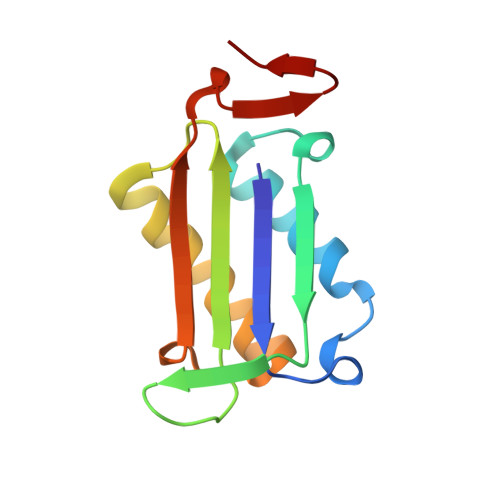
Inactivation of the phenylpyruvate tautomerase activity of macrophage migration inhibitory factor by 2-oxo-4-phenyl-3-butynoate.
Golubkov, P.A., Johnson Jr., W.H., Czerwinski, R.M., Person, M.D., Wang, S.C., Whitman, C.P., Hackert, M.L.(2006) Bioorg Chem 34: 183-199
- PubMed: 16780921
- DOI: https://doi.org/10.1016/j.bioorg.2006.05.001
- Primary Citation of Related Structures:
2GDG - PubMed Abstract:
Macrophage migration inhibitory factor (MIF) is an important immunoregulatory protein that has been implicated in several inflammatory diseases. MIF also has a phenylpyruvate tautomerase (PPT) activity, the role of which remains elusive in these biological activities. The acetylene compound, 2-oxo-4-phenyl-3-butynoate (2-OPB), has been synthesized and tested as a potential irreversible inhibitor of its enzymatic activity. Incubation of the compound with MIF results in the rapid and irreversible loss of the PPT activity. Mass spectral analysis established that the amino-terminal proline, previously implicated as a catalytic base in the PPT-catalyzed reaction, is the site of covalent modification. Inactivation of the PPT activity likely occurs by a Michael addition of Pro-1 to C-4 of the inhibitor. Attempts to crystallize the inactivated complex to confirm the structure of the adduct on the covalently modified Pro-1 by X-ray crystallography were not successful. Nor was it possible to unambiguously interpret electron density observed in the active sites of the native crystals soaked with the inhibitor. This may be due to crystal packing in that the side chain of Glu-16 from an adjacent trimer occupies one active site. However, this crystal contact may be partially responsible for the high-resolution quality of these MIF crystals. Nonetheless, because MIF is a member of the tautomerase superfamily, a group of structurally homologous proteins that share a beta-alpha-beta structural motif and a catalytic Pro-1, 2-OPB may find general use as a probe of tautomerase superfamily members that function as PPTs.
Organizational Affiliation:
Department of Chemistry and Biochemistry, University of Texas at Austin, Austin, TX 78712, USA.


