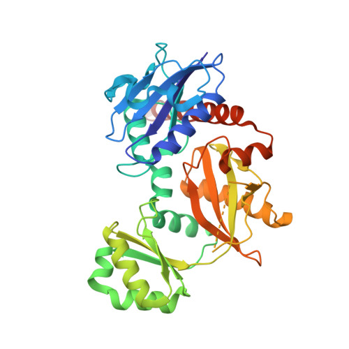
Allosteric inhibition of Staphylococcus aureus D-alanine:D-alanine ligase revealed by crystallographic studies.
Liu, S., Chang, J.S., Herberg, J.T., Horng, M.M., Tomich, P.K., Lin, A.H., Marotti, K.R.(2006) Proc Natl Acad Sci U S A 103: 15178-15183
- PubMed: 17015835
- DOI: https://doi.org/10.1073/pnas.0604905103
- Primary Citation of Related Structures:
2I80, 2I87, 2I8C - PubMed Abstract:
D-alanine:D-alanine ligase (DDl) is an essential enzyme in bacterial cell wall biosynthesis and an important target for developing new antibiotics. It catalyzes the formation of D-alanine:D-alanine dipeptide, sequentially by using one D-alanine and one ATP as substrates for the first-half reaction, and a second D-alanine substrate to complete the reaction. Some gain of function DDl mutants can use an alternate second substrate, causing resistance to vancomycin, one of the last lines of defense against life-threatening Gram-positive infections. Here, we report the crystal structure of Staphylococcus aureus DDl (StaDDl) and its cocrystal structures with 3-chloro-2,2-dimethyl-N-[4(trifluoromethyl)phenyl]propanamide (inhibitor 1) (Ki=4 microM against StaDDl) and with ADP, one of the reaction products, at resolutions of 2.0, 2.2, and 2.6 A, respectively. The overall structure of StaDDl can be divided into three distinct domains. The inhibitor binds to a hydrophobic pocket at the interface of the first and the third domain. This inhibitor-binding pocket is adjacent to the first D-alanine substrate site but does not overlap with any substrate sites. An allosteric inhibition mechanism of StaDDl by this compound was proposed. The mechanism provides the basis for developing new antibiotics targeting D-alanine:D-alanine ligase. Because this compound only interacts with residues from the first D-alanine site, inhibitors with this binding mode potentially could overcome vancomycin resistance.
Organizational Affiliation:
Pfizer, Inc., Eastern Point Road, Groton, CT 06340, USA. [email protected]


