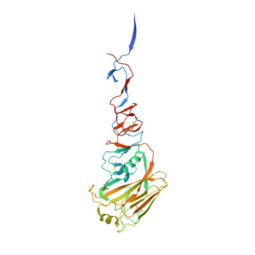
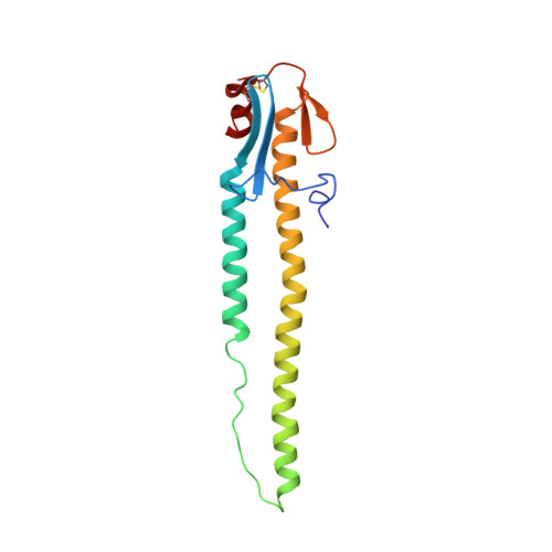
2IBX

Influenza virus (VN1194) H5 HA


Experimental Data Snapshot

  • Method: X-RAY DIFFRACTION
  • Resolution: 2.80 Å
  • R-Value Free: 0.302 
  • R-Value Work: 0.241 
  • R-Value Observed: 0.244 

Starting Model: experimental
View more details

wwPDB Validation   3D Report Full Report


Ligand Structure Quality Assessment 


This is version 2.1 of the entry. See complete history


Literature

Haemagglutinin mutations responsible for the binding of H5N1 influenza A viruses to human-type receptors.

Yamada, S.Suzuki, Y.Suzuki, T.Le, M.Q.Nidom, C.A.Sakai-Tagawa, Y.Muramoto, Y.Ito, M.Kiso, M.Horimoto, T.Shinya, K.Sawada, T.Kiso, M.Usui, T.Murata, T.Lin, Y.Hay, A.Haire, L.F.Stevens, D.J.Russell, R.J.Gamblin, S.J.Skehel, J.J.Kawaoka, Y.

(2006) Nature 444: 378-382

  • DOI: https://doi.org/10.1038/nature05264
  • Primary Citation of Related Structures:  
    2IBX

  • PubMed Abstract: 

    H5N1 influenza A viruses have spread to numerous countries in Asia, Europe and Africa, infecting not only large numbers of poultry, but also an increasing number of humans, often with lethal effects. Human and avian influenza A viruses differ in their recognition of host cell receptors: the former preferentially recognize receptors with saccharides terminating in sialic acid-alpha2,6-galactose (SAalpha2,6Gal), whereas the latter prefer those ending in SAalpha2,3Gal (refs 3-6). A conversion from SAalpha2,3Gal to SAalpha2,6Gal recognition is thought to be one of the changes that must occur before avian influenza viruses can replicate efficiently in humans and acquire the potential to cause a pandemic. By identifying mutations in the receptor-binding haemagglutinin (HA) molecule that would enable avian H5N1 viruses to recognize human-type host cell receptors, it may be possible to predict (and thus to increase preparedness for) the emergence of pandemic viruses. Here we show that some H5N1 viruses isolated from humans can bind to both human and avian receptors, in contrast to those isolated from chickens and ducks, which recognize the avian receptors exclusively. Mutations at positions 182 and 192 independently convert the HAs of H5N1 viruses known to recognize the avian receptor to ones that recognize the human receptor. Analysis of the crystal structure of the HA from an H5N1 virus used in our genetic experiments shows that the locations of these amino acids in the HA molecule are compatible with an effect on receptor binding. The amino acid changes that we identify might serve as molecular markers for assessing the pandemic potential of H5N1 field isolates.


  • Organizational Affiliation

    Division of Virology, Department of Microbiology and Immunology, Institute of Medical Science, University of Tokyo, Tokyo 108-8639, Japan.


Macromolecules
Find similar proteins by:  (by identity cutoff)  |  3D Structure
Entity ID: 1
MoleculeChains Sequence LengthOrganismDetailsImage
Hemagglutinin
A, C, E
340Influenza A virusMutation(s): 0 
Gene Names: HA
UniProt
Find proteins for Q45ZQ3 (Influenza A virus)
Explore Q45ZQ3 
Go to UniProtKB:  Q45ZQ3
Entity Groups  
Sequence Clusters30% Identity50% Identity70% Identity90% Identity95% Identity100% Identity
UniProt GroupQ45ZQ3
Glycosylation
Glycosylation Sites: 2
Sequence Annotations
Expand
  • Reference Sequence
Find similar proteins by:  (by identity cutoff)  |  3D Structure
Entity ID: 2
MoleculeChains Sequence LengthOrganismDetailsImage
Hemagglutinin
B, D, F
160Influenza A virusMutation(s): 0 
Gene Names: HA
UniProt
Find proteins for Q66QC3 (Influenza A virus)
Explore Q66QC3 
Go to UniProtKB:  Q66QC3
Entity Groups  
Sequence Clusters30% Identity50% Identity70% Identity90% Identity95% Identity100% Identity
UniProt GroupQ66QC3
Sequence Annotations
Expand
  • Reference Sequence
Oligosaccharides

Help

Entity ID: 3
MoleculeChains Length2D Diagram Glycosylation3D Interactions
2-acetamido-2-deoxy-beta-D-glucopyranose-(1-4)-2-acetamido-2-deoxy-beta-D-glucopyranose
G, H, I
2N-Glycosylation
Glycosylation Resources
GlyTouCan:  G42666HT
GlyCosmos:  G42666HT
GlyGen:  G42666HT
Experimental Data & Validation

Experimental Data

  • Method: X-RAY DIFFRACTION
  • Resolution: 2.80 Å
  • R-Value Free: 0.302 
  • R-Value Work: 0.241 
  • R-Value Observed: 0.244 
  • Space Group: C 1 2 1
Unit Cell:
Length ( Å )Angle ( ˚ )
a = 174.678α = 90
b = 100.563β = 111.39
c = 160.13γ = 90
Software Package:
Software NamePurpose
REFMACrefinement
DENZOdata reduction
SCALEPACKdata scaling
AMoREphasing

Structure Validation

View Full Validation Report


Ligand Structure Quality Assessment 


Entry History 

Deposition Data

Revision History  (Full details and data files)

  • Version 1.0: 2006-11-28
    Type: Initial release
  • Version 1.1: 2008-05-01
    Changes: Version format compliance
  • Version 1.2: 2011-07-13
    Changes: Non-polymer description, Version format compliance
  • Version 2.0: 2020-07-29
    Type: Remediation
    Reason: Carbohydrate remediation
    Changes: Advisory, Atomic model, Data collection, Derived calculations, Structure summary
  • Version 2.1: 2023-08-30
    Changes: Data collection, Database references, Refinement description, Structure summary