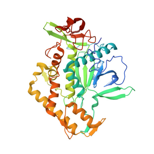
2IMB

Clostridium botulinum Neurotoxin Serotype A Light Chain Inhibited by L-arginine hydroxamate


Experimental Data Snapshot

  • Method: X-RAY DIFFRACTION
  • Resolution: 2.41 Å
  • R-Value Free: 0.260 
  • R-Value Work: 0.191 
  • R-Value Observed: 0.195 

wwPDB Validation   3D Report Full Report


Ligand Structure Quality Assessment 


This is version 1.4 of the entry. See complete history


Literature

Structures of Clostridium botulinum Neurotoxin Serotype A Light Chain Complexed with Small-Molecule Inhibitors Highlight Active-Site Flexibility.

Silvaggi, N.R.Boldt, G.E.Hixon, M.S.Kennedy, J.P.Tzipori, S.Janda, K.D.Allen, K.N.

(2007) Chem Biol 14: 533-542

  • DOI: https://doi.org/10.1016/j.chembiol.2007.03.014
  • Primary Citation of Related Structures:  
    2ILP, 2IMA, 2IMB, 2IMC

  • PubMed Abstract: 

    The potential for the use of Clostridial neurotoxins as bioweapons makes the development of small-molecule inhibitors of these deadly toxins a top priority. Recently, screening of a random hydroxamate library identified a small-molecule inhibitor of C. botulinum Neurotoxin Serotype A Light Chain (BoNT/A-LC), 4-chlorocinnamic hydroxamate, a derivative of which has been shown to have in vivo efficacy in mice and no toxicity. We describe the X-ray crystal structures of BoNT/A-LC in complexes with two potent small-molecule inhibitors. The structures of the enzyme with 4-chlorocinnamic hydroxamate or 2,4-dichlorocinnamic hydroxamate bound are compared to the structure of the enzyme complexed with L-arginine hydroxamate, an inhibitor with modest affinity. Taken together, this suite of structures provides surprising insights into the BoNT/A-LC active site, including unexpected conformational flexibility at the S1' site that changes the electrostatic environment of the binding pocket. Information gained from these structures will inform the design and optimization of more effective small-molecule inhibitors of BoNT/A-LC.


  • Organizational Affiliation

    Department of Physiology and Biophysics, Boston University School of Medicine, Boston, MA 02118, USA.


Macromolecules
Find similar proteins by:  (by identity cutoff)  |  3D Structure
Entity ID: 1
MoleculeChains Sequence LengthOrganismDetailsImage
Botulinum neurotoxin A light-chain
A, B
444Clostridium botulinumMutation(s): 0 
Gene Names: botAatxbna
EC: 3.4.24.69
UniProt
Find proteins for P0DPI1 (Clostridium botulinum (strain Hall / ATCC 3502 / NCTC 13319 / Type A))
Explore P0DPI1 
Go to UniProtKB:  P0DPI1
Entity Groups  
Sequence Clusters30% Identity50% Identity70% Identity90% Identity95% Identity100% Identity
UniProt GroupP0DPI1
Sequence Annotations
Expand
  • Reference Sequence
Experimental Data & Validation

Experimental Data

  • Method: X-RAY DIFFRACTION
  • Resolution: 2.41 Å
  • R-Value Free: 0.260 
  • R-Value Work: 0.191 
  • R-Value Observed: 0.195 
  • Space Group: P 1 21 1
Unit Cell:
Length ( Å )Angle ( ˚ )
a = 74.238α = 90
b = 67.928β = 105.1
c = 98.305γ = 90
Software Package:
Software NamePurpose
DENZOdata reduction
SCALEPACKdata scaling
REFMACrefinement
PDB_EXTRACTdata extraction
CBASSdata collection

Structure Validation

View Full Validation Report


Ligand Structure Quality Assessment 


Entry History 

Deposition Data

Revision History  (Full details and data files)

  • Version 1.0: 2007-06-05
    Type: Initial release
  • Version 1.1: 2008-05-01
    Changes: Version format compliance
  • Version 1.2: 2011-07-13
    Changes: Version format compliance
  • Version 1.3: 2017-10-18
    Changes: Refinement description
  • Version 1.4: 2024-02-21
    Changes: Data collection, Database references, Derived calculations