2J17

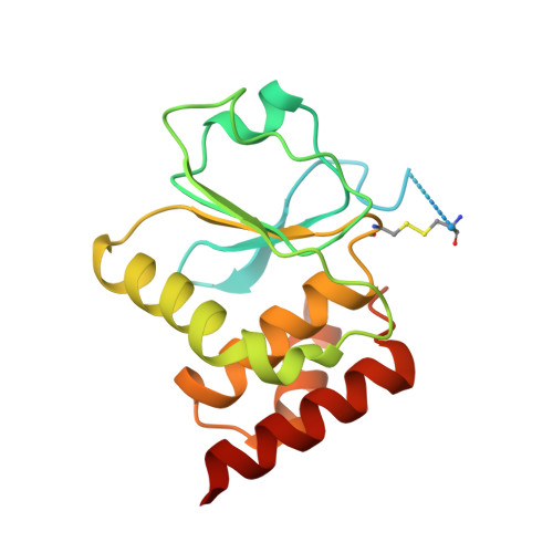
pTyr bound form of SDP-1


Experimental Data Snapshot

  • Method: X-RAY DIFFRACTION
  • Resolution: 2.84 Å
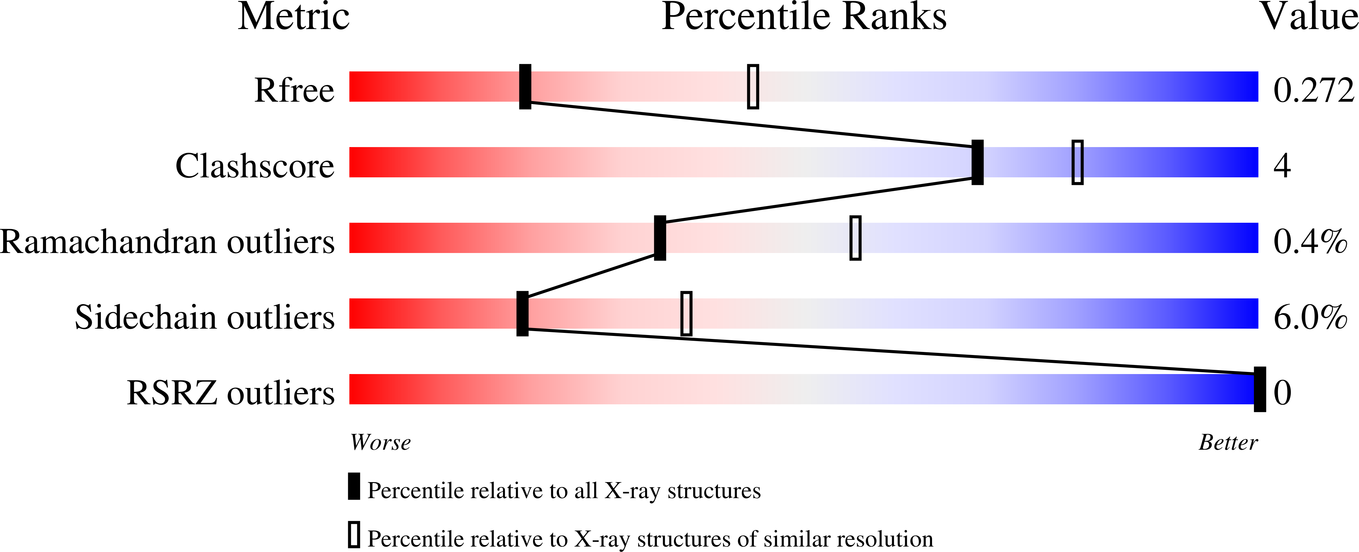
  • R-Value Free: 0.269 
  • R-Value Work: 0.221 
  • R-Value Observed: 0.224 

Starting Model: experimental
View more details

wwPDB Validation   3D Report Full Report


Ligand Structure Quality Assessment 


This is version 1.6 of the entry. See complete history


Literature

Redox-mediated substrate recognition by Sdp1 defines a new group of tyrosine phosphatases.

Fox, G.C.Shafiq, M.Briggs, D.C.Knowles, P.P.Collister, M.Didmon, M.J.Makrantoni, V.Dickinson, R.J.Hanrahan, S.Totty, N.Stark, M.J.Keyse, S.M.McDonald, N.Q.

(2007) Nature 447: 487-492

  • DOI: https://doi.org/10.1038/nature05804
  • Primary Citation of Related Structures:  
    2J16, 2J17

  • PubMed Abstract: 

    Reactive oxygen species trigger cellular responses by activation of stress-responsive mitogen-activated protein kinase (MAPK) signalling pathways. Reversal of MAPK activation requires the transcriptional induction of specialized cysteine-based phosphatases that mediate MAPK dephosphorylation. Paradoxically, oxidative stresses generally inactivate cysteine-based phosphatases by thiol modification and thus could lead to sustained or uncontrolled MAPK activation. Here we describe how the stress-inducible MAPK phosphatase, Sdp1, presents an unusual solution to this apparent paradox by acquiring enhanced catalytic activity under oxidative conditions. Structural and biochemical evidence reveals that Sdp1 employs an intramolecular disulphide bridge and an invariant histidine side chain to selectively recognize a tyrosine-phosphorylated MAPK substrate. Optimal activity critically requires the disulphide bridge, and thus, to the best of our knowledge, Sdp1 is the first example of a cysteine-dependent phosphatase that couples oxidative stress with substrate recognition. We show that Sdp1, and its paralogue Msg5, have similar properties and belong to a new group of phosphatases unique to yeast and fungal taxa.


  • Organizational Affiliation

    Structural Biology Laboratory, The London Research Institute, Cancer Research UK, 44 Lincoln's Inn Fields, London WC2A 3PX, UK.


Macromolecules
Find similar proteins by:  (by identity cutoff)  |  3D Structure
Entity ID: 1
MoleculeChains Sequence LengthOrganismDetailsImage
TYROSINE-PROTEIN PHOSPHATASE YIL113W
A, B
182Saccharomyces cerevisiaeMutation(s): 1 
EC: 3.1.3.48
UniProt
Find proteins for P40479 (Saccharomyces cerevisiae (strain ATCC 204508 / S288c))
Explore P40479 
Go to UniProtKB:  P40479
Entity Groups  
Sequence Clusters30% Identity50% Identity70% Identity90% Identity95% Identity100% Identity
UniProt GroupP40479
Sequence Annotations
Expand
  • Reference Sequence
Experimental Data & Validation

Experimental Data

  • Method: X-RAY DIFFRACTION
  • Resolution: 2.84 Å
  • R-Value Free: 0.269 
  • R-Value Work: 0.221 
  • R-Value Observed: 0.224 
  • Space Group: P 43 21 2
Unit Cell:
Length ( Å )Angle ( ˚ )
a = 61.661α = 90
b = 61.661β = 90
c = 217.562γ = 90
Software Package:
Software NamePurpose
REFMACrefinement
MOSFLMdata reduction
SCALAdata scaling
MOLREPphasing

Structure Validation

View Full Validation Report


Ligand Structure Quality Assessment 


Entry History 

Deposition Data

Revision History  (Full details and data files)

  • Version 1.0: 2007-05-22
    Type: Initial release
  • Version 1.1: 2011-05-08
    Changes: Version format compliance
  • Version 1.2: 2011-07-13
    Changes: Version format compliance
  • Version 1.3: 2018-02-28
    Changes: Database references
  • Version 1.4: 2019-05-08
    Changes: Data collection, Experimental preparation
  • Version 1.5: 2023-12-13
    Changes: Data collection, Database references, Derived calculations, Other, Refinement description
  • Version 1.6: 2024-11-06
    Changes: Structure summary