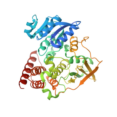
Structures of Mycobacterium Tuberculosis 1-Deoxy-D- Xylulose-5-Phosphate Reductoisomerase Provide New Insights Into Catalysis.
Henriksson, L.M., Unge, T., Carlsson, J., Aqvist, J., Mowbray, S.L., Jones, T.A.(2007) J Biol Chem 282: 19905
- PubMed: 17491006
- DOI: https://doi.org/10.1074/jbc.M701935200
- Primary Citation of Related Structures:
2JCV, 2JCX, 2JCY, 2JD0, 2JD1, 2JD2, 4AIC - PubMed Abstract:
Isopentenyl diphosphate is the precursor of various isoprenoids that are essential to all living organisms. It is produced by the mevalonate pathway in humans but by an alternate route in plants, protozoa, and many bacteria. 1-deoxy-D-xylulose-5-phosphate reductoisomerase catalyzes the second step of this non-mevalonate pathway, which involves an NADPH-dependent rearrangement and reduction of 1-deoxy-D-xylulose 5-phosphate to form 2-C-methyl-D-erythritol 4-phosphate. The use of different pathways, combined with the reported essentiality of the enzyme makes the reductoisomerase a highly promising target for drug design. Here we present several high resolution structures of the Mycobacterium tuberculosis 1-deoxy-D-xylulose-5-phosphate reductoisomerase, representing both wild type and mutant enzyme in various complexes with Mn(2+), NADPH, and the known inhibitor fosmidomycin. The asymmetric unit corresponds to the biological homodimer. Although crystal contacts stabilize an open active site in the B molecule, the A molecule displays a closed conformation, with some differences depending on the ligands bound. An inhibition study with fosmidomycin resulted in an estimated IC(50) value of 80 nm. The double mutant enzyme (D151N/E222Q) has lost its ability to bind the metal and, thereby, also its activity. Our structural information complemented with molecular dynamics simulations and free energy calculations provides the framework for the design of new inhibitors and gives new insights into the reaction mechanism. The conformation of fosmidomycin bound to the metal ion is different from that reported in a previously published structure and indicates that a rearrangement of the intermediate is not required during catalysis.
Organizational Affiliation:
Department of Cell and Molecular Biology, Uppsala University, Biomedical Center, Box 596, SE-751 24 Uppsala, Sweden.


