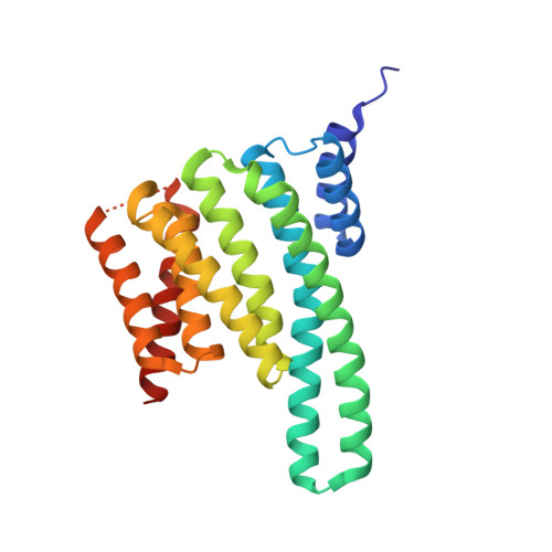
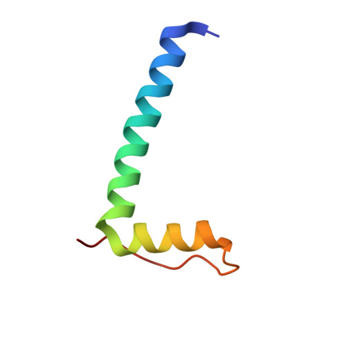
2O98

Structure of the 14-3-3 / H+-ATPase plant complex


Experimental Data Snapshot

  • Method: X-RAY DIFFRACTION
  • Resolution: 2.70 Å
  • R-Value Free: 0.263 
  • R-Value Work: 0.178 
  • R-Value Observed: 0.182 

wwPDB Validation   3D Report Full Report


Ligand Structure Quality Assessment 


This is version 1.5 of the entry. See complete history


Literature

Structure of a 14-3-3 coordinated hexamer of the plant plasma membrane H+ -ATPase by combining X-ray crystallography and electron cryomicroscopy

Ottmann, C.Marco, S.Jaspert, N.Marcon, C.Schauer, N.Weyand, M.Vandermeeren, C.Duby, G.Boutry, M.Wittinghofer, A.Rigaud, J.L.Oecking, C.

(2007) Mol Cell 25: 427-440

  • DOI: https://doi.org/10.1016/j.molcel.2006.12.017
  • Primary Citation of Related Structures:  
    2O98

  • PubMed Abstract: 

    Regulatory 14-3-3 proteins activate the plant plasma membrane H(+)-ATPase by binding to its C-terminal autoinhibitory domain. This interaction requires phosphorylation of a C-terminal, mode III, recognition motif as well as an adjacent span of approximately 50 amino acids. Here we report the X-ray crystal structure of 14-3-3 in complex with the entire binding motif, revealing a previously unidentified mode of interaction. A 14-3-3 dimer simultaneously binds two H(+)-ATPase peptides, each of which forms a loop within the typical 14-3-3 binding groove and therefore exits from the center of the dimer. Several H(+)-ATPase mutants support this structure determination. Accordingly, 14-3-3 binding could result in H(+)-ATPase oligomerization. Indeed, by using single-particle electron cryomicroscopy, the 3D reconstruction of the purified H(+)-ATPase/14-3-3 complex demonstrates a hexameric arrangement. Fitting of 14-3-3 and H(+)-ATPase atomic structures into the 3D reconstruction map suggests the spatial arrangement of the holocomplex.


  • Organizational Affiliation

    Zentrum für Molekularbiologie der Pflanzen, Pflanzenphysiologie, Universität Tübingen, Auf der Morgenstelle 5, 72076 Tübingen, Germany.


Macromolecules
Find similar proteins by:  (by identity cutoff)  |  3D Structure
Entity ID: 1
MoleculeChains Sequence LengthOrganismDetailsImage
14-3-3-like protein C
A, B
242Nicotiana tabacumMutation(s): 0 
UniProt
Find proteins for P93343 (Nicotiana tabacum)
Explore P93343 
Go to UniProtKB:  P93343
Entity Groups  
Sequence Clusters30% Identity50% Identity70% Identity90% Identity95% Identity100% Identity
UniProt GroupP93343
Sequence Annotations
Expand
  • Reference Sequence
Find similar proteins by:  (by identity cutoff)  |  3D Structure
Entity ID: 2
MoleculeChains Sequence LengthOrganismDetailsImage
Plasma membrane H+ ATPaseC [auth P],
D [auth Q]
52Nicotiana plumbaginifoliaMutation(s): 2 
Gene Names: pma2
EC: 7.1.2.1
UniProt
Find proteins for Q08436 (Nicotiana plumbaginifolia)
Explore Q08436 
Go to UniProtKB:  Q08436
Entity Groups  
Sequence Clusters30% Identity50% Identity70% Identity90% Identity95% Identity100% Identity
UniProt GroupQ08436
Sequence Annotations
Expand
  • Reference Sequence
Experimental Data & Validation

Experimental Data

  • Method: X-RAY DIFFRACTION
  • Resolution: 2.70 Å
  • R-Value Free: 0.263 
  • R-Value Work: 0.178 
  • R-Value Observed: 0.182 
  • Space Group: I 41 2 2
Unit Cell:
Length ( Å )Angle ( ˚ )
a = 114.37α = 90
b = 114.37β = 90
c = 236.79γ = 90
Software Package:
Software NamePurpose
REFMACrefinement
PDB_EXTRACTdata extraction
MAR345data collection
XDSdata reduction
XSCALEdata scaling
AMoREphasing

Structure Validation

View Full Validation Report


Ligand Structure Quality Assessment 


Entry History 

Deposition Data

Revision History  (Full details and data files)

  • Version 1.0: 2007-04-03
    Type: Initial release
  • Version 1.1: 2008-05-01
    Changes: Version format compliance
  • Version 1.2: 2011-07-13
    Changes: Advisory, Version format compliance
  • Version 1.3: 2017-10-18
    Changes: Advisory, Refinement description
  • Version 1.4: 2021-11-10
    Changes: Advisory, Database references, Derived calculations
  • Version 1.5: 2023-12-27
    Changes: Data collection