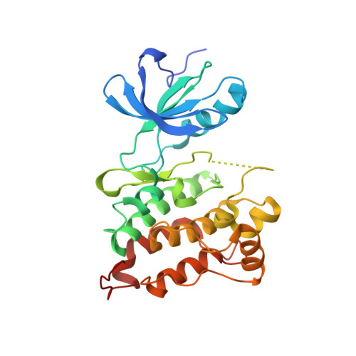
2OIQ

Crystal Structure of chicken c-Src kinase domain in complex with the cancer drug imatinib.


Experimental Data Snapshot

  • Method: X-RAY DIFFRACTION
  • Resolution: 2.07 Å
  • R-Value Free: 0.263 
  • R-Value Work: 0.227 
  • R-Value Observed: 0.232 

Starting Model: experimental
View more details

wwPDB Validation   3D Report Full Report


Ligand Structure Quality Assessment 


This is version 1.4 of the entry. See complete history


Literature

c-Src Binds to the Cancer Drug Imatinib with an Inactive Abl/c-Kit Conformation and a Distributed Thermodynamic Penalty.

Seeliger, M.A.Nagar, B.Frank, F.Cao, X.Henderson, M.N.Kuriyan, J.

(2007) Structure 15: 299-311

  • DOI: https://doi.org/10.1016/j.str.2007.01.015
  • Primary Citation of Related Structures:  
    2OIQ

  • PubMed Abstract: 

    The cancer drug imatinib inhibits the tyrosine kinases c-Abl, c-Kit, and the PDGF receptor. Imatinib is less effective against c-Src, which is difficult to understand because residues interacting with imatinib in crystal structures of Abl and c-Kit are conserved in c-Src. The crystal structure of the c-Src kinase domain in complex with imatinib closely resembles that of Abl*imatinib and c-Kit*imatinib, and differs significantly from the inactive "Src/CDK" conformation of the Src family kinases. Attempts to increase the affinity of c-Src for imatinib by swapping residues with the corresponding residues in Abl have not been successful, suggesting that the thermodynamic penalty for adoption of the imatinib-binding conformation by c-Src is distributed over a broad region of the structure. Two mutations that are expected to destabilize the inactive Src/CDK conformation increase drug sensitivity 15-fold, suggesting that the free-energy balance between different inactive states is a key to imatinib binding.


  • Organizational Affiliation

    Howard Hughes Medical Institute, University of California, Berkeley, Berkeley, CA 94720, USA.


Macromolecules
Find similar proteins by:  (by identity cutoff)  |  3D Structure
Entity ID: 1
MoleculeChains Sequence LengthOrganismDetailsImage
Proto-oncogene tyrosine-protein kinase Src
A, B
286Gallus gallusMutation(s): 0 
Gene Names: SRC
EC: 2.7.10.2
UniProt
Find proteins for P00523 (Gallus gallus)
Explore P00523 
Go to UniProtKB:  P00523
Entity Groups  
Sequence Clusters30% Identity50% Identity70% Identity90% Identity95% Identity100% Identity
UniProt GroupP00523
Sequence Annotations
Expand
  • Reference Sequence
Small Molecules
Binding Affinity Annotations 
IDSourceBinding Affinity
STI BindingDB:  2OIQ Ki: min: 1.00e+4, max: 3.10e+4 (nM) from 2 assay(s)
Kd: min: 1.00e+4, max: 3.98e+4 (nM) from 3 assay(s)
IC50: min: 2784, max: 1.00e+5 (nM) from 3 assay(s)
PDBBind:  2OIQ Kd: 1.00e+4 (nM) from 1 assay(s)
Experimental Data & Validation

Experimental Data

  • Method: X-RAY DIFFRACTION
  • Resolution: 2.07 Å
  • R-Value Free: 0.263 
  • R-Value Work: 0.227 
  • R-Value Observed: 0.232 
  • Space Group: P 1
Unit Cell:
Length ( Å )Angle ( ˚ )
a = 42.078α = 78.99
b = 63.373β = 89.12
c = 74.474γ = 90.1
Software Package:
Software NamePurpose
HKL-2000data collection
PHASERphasing
CNSrefinement
DENZOdata reduction
SCALEPACKdata scaling

Structure Validation

View Full Validation Report


Ligand Structure Quality Assessment 


Entry History 

Deposition Data

Revision History  (Full details and data files)

  • Version 1.0: 2007-03-20
    Type: Initial release
  • Version 1.1: 2008-05-01
    Changes: Version format compliance
  • Version 1.2: 2011-07-13
    Changes: Non-polymer description, Version format compliance
  • Version 1.3: 2020-04-15
    Changes: Data collection, Database references
  • Version 1.4: 2023-08-30
    Changes: Data collection, Database references, Derived calculations, Refinement description, Structure summary