2PEG

Crystal structure of Trematomus bernacchii hemoglobin in a partial hemichrome state


Experimental Data Snapshot

  • Method: X-RAY DIFFRACTION
  • Resolution: 1.48 Å
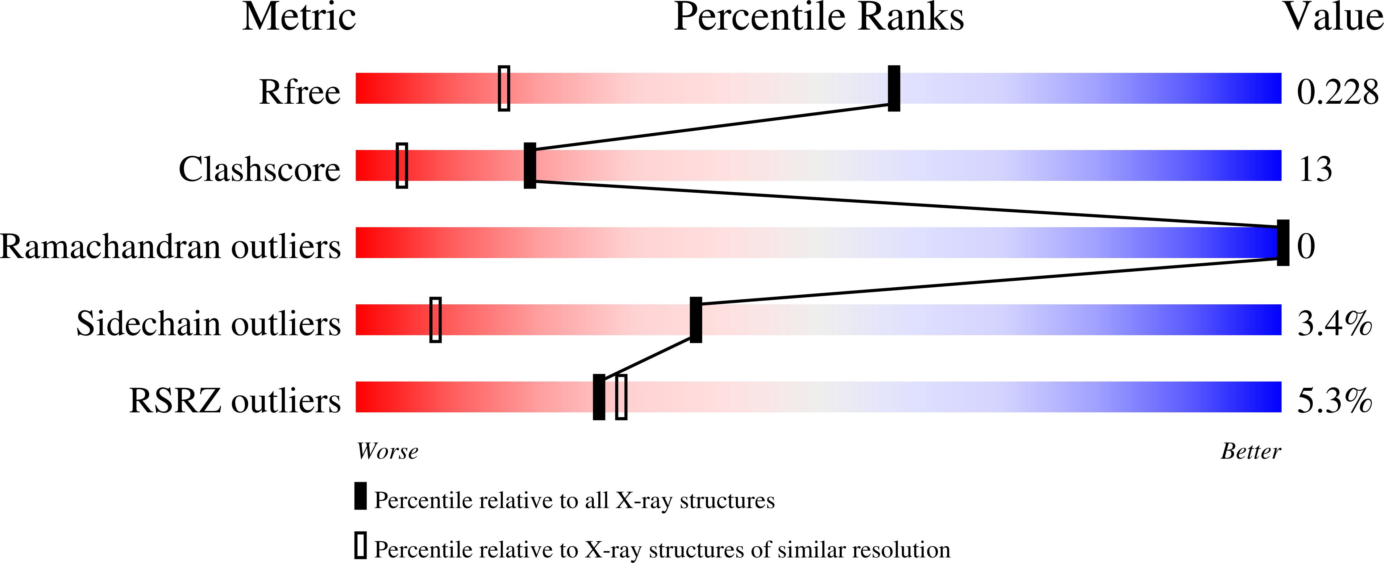
  • R-Value Free: 0.234 
  • R-Value Work: 0.199 
  • R-Value Observed: 0.203 

Starting Model: experimental
View more details

wwPDB Validation   3D Report Full Report


Ligand Structure Quality Assessment 


This is version 1.6 of the entry. See complete history


Literature

Structural characterization of ferric hemoglobins from three antarctic fish species of the suborder notothenioidei.

Vergara, A.Franzese, M.Merlino, A.Vitagliano, L.Verde, C.di Prisco, G.Lee, H.C.Peisach, J.Mazzarella, L.

(2007) Biophys J 93: 2822-2829

  • DOI: https://doi.org/10.1529/biophysj.107.105700
  • Primary Citation of Related Structures:  
    2PEG

  • PubMed Abstract: 

    Spontaneous autoxidation of tetrameric Hbs leads to the formation of Fe (III) forms, whose physiological role is not fully understood. Here we report structural characterization by EPR of the oxidized states of tetrameric Hbs isolated from the Antarctic fish species Trematomus bernacchii, Trematomus newnesi, and Gymnodraco acuticeps, as well as the x-ray crystal structure of oxidized Trematomus bernacchii Hb, redetermined at high resolution. The oxidation of these Hbs leads to formation of states that were not usually detected in previous analyses of tetrameric Hbs. In addition to the commonly found aquo-met and hydroxy-met species, EPR analyses show that two distinct hemichromes coexist at physiological pH, referred to as hemichromes I and II, respectively. Together with the high-resolution crystal structure (1.5 A) of T. bernacchii and a survey of data available for other heme proteins, hemichrome I was assigned by x-ray crystallography and by EPR as a bis-His complex with a distorted geometry, whereas hemichrome II is a less constrained (cytochrome b5-like) bis-His complex. In four of the five Antartic fish Hbs examined, hemichrome I is the major form. EPR shows that for HbCTn, the amount of hemichrome I is substantially reduced. In addition, the concomitant presence of a penta-coordinated high-spin Fe (III) species, to our knowledge never reported before for a wild-type tetrameric Hb, was detected. A molecular modeling investigation demonstrates that the presence of the bulkier Ile in position 67beta in HbCTn in place of Val as in the other four Hbs impairs the formation of hemichrome I, thus favoring the formation of the ferric penta-coordinated species. Altogether the data show that ferric states commonly associated with monomeric and dimeric Hbs are also found in tetrameric Hbs.


  • Organizational Affiliation

    Department of Chemistry, University of Naples Federico II, Complesso Universitario Monte S. Angelo, I-80126 Naples, Italy.


Macromolecules
Find similar proteins by:  (by identity cutoff)  |  3D Structure
Entity ID: 1
MoleculeChains Sequence LengthOrganismDetailsImage
Hemoglobin subunit alpha143Trematomus bernacchiiMutation(s): 0 
UniProt
Find proteins for P80043 (Trematomus bernacchii)
Explore P80043 
Go to UniProtKB:  P80043
Entity Groups  
Sequence Clusters30% Identity50% Identity70% Identity90% Identity95% Identity100% Identity
UniProt GroupP80043
Sequence Annotations
Expand
  • Reference Sequence
Find similar proteins by:  (by identity cutoff)  |  3D Structure
Entity ID: 2
MoleculeChains Sequence LengthOrganismDetailsImage
Hemoglobin subunit beta146Trematomus bernacchiiMutation(s): 0 
UniProt
Find proteins for P80044 (Trematomus bernacchii)
Explore P80044 
Go to UniProtKB:  P80044
Entity Groups  
Sequence Clusters30% Identity50% Identity70% Identity90% Identity95% Identity100% Identity
UniProt GroupP80044
Sequence Annotations
Expand
  • Reference Sequence
Experimental Data & Validation

Experimental Data

  • Method: X-RAY DIFFRACTION
  • Resolution: 1.48 Å
  • R-Value Free: 0.234 
  • R-Value Work: 0.199 
  • R-Value Observed: 0.203 
  • Space Group: C 1 2 1
Unit Cell:
Length ( Å )Angle ( ˚ )
a = 87.16α = 90
b = 87.74β = 97.66
c = 55.38γ = 90
Software Package:
Software NamePurpose
X-PLORrefinement
SCALEPACKdata scaling
CNSrefinement
PDB_EXTRACTdata extraction
MAR345dtbdata collection
AMoREphasing
DENZOdata reduction

Structure Validation

View Full Validation Report


Ligand Structure Quality Assessment 


Entry History 

Deposition Data

Revision History  (Full details and data files)

  • Version 1.0: 2007-07-24
    Type: Initial release
  • Version 1.1: 2007-10-09
    Changes: Version format compliance
  • Version 1.2: 2011-07-13
    Changes: Version format compliance
  • Version 1.3: 2017-10-18
    Changes: Refinement description
  • Version 1.4: 2019-07-24
    Changes: Data collection, Derived calculations, Refinement description
  • Version 1.5: 2023-08-30
    Changes: Data collection, Database references, Derived calculations, Refinement description
  • Version 1.6: 2024-10-09
    Changes: Structure summary