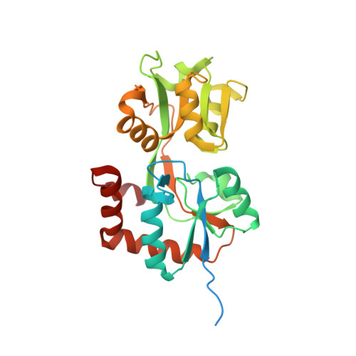
Crystal structures and mutational analysis of the arginine-, lysine-, histidine-binding protein ArtJ from Geobacillus stearothermophilus. Implications for interactions of ArtJ with its cognate ATP-binding cassette transporter, Art(MP)2
Vahedi-Faridi, A., Eckey, V., Scheffel, F., Alings, C., Landmesser, H., Schneider, E., Saenger, W.(2008) J Mol Biol 375: 448-459
- PubMed: 18022195
- DOI: https://doi.org/10.1016/j.jmb.2007.10.049
- Primary Citation of Related Structures:
2PVU, 2Q2A, 2Q2C - PubMed Abstract:
ArtJ is the substrate-binding component (receptor) of the ATP-binding cassette (ABC) transport system ArtJ-(MP)(2) from the thermophilic bacterium Geobacillus stearothermophilus that is specific for arginine, lysine, and histidine. The highest affinity is found for arginine (K(d)=0.039(+/-0.014) microM), while the affinities for lysine and histidine are about tenfold lower. We have determined the X-ray structures of ArtJ liganded with each of these substrates at resolutions of 1.79 A (arginine), 1.79 A (lysine), and 2.35 A (histidine), respectively. As found for other solute receptors, the polypeptide chain is folded into two distinct domains (lobes) connected by a hinge. The interface between the lobes forms the substrate-binding pocket whose geometry is well preserved in all three ArtJ/amino acid complexes. Structure-derived mutational analyses indicated the crucial role of a region in the carboxy-terminal lobe of ArtJ in contacting the transport pore Art(MP)(2) and revealed the functional importance of Gln132 and Trp68. While variant Gln132Leu exhibited lower binding affinity for arginine but no binding of lysine and histidine, the variant Trp68Leu had lost binding activity for all three substrates. The results are discussed in comparison with known structures of homologous proteins from mesophilic bacteria.
Organizational Affiliation:
Institut für Chemie und Biochemie/Kristallographie, Freie Universitaet Berlin, Takustr. 6, 14195 Berlin, Germany.


