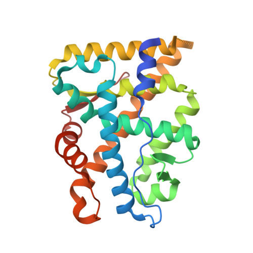
2Q7K

The Androgen Receptor Prostate Cancer Mutant H874Y Ligand Binding Domain Bound with Testosterone and an AR 20-30 Peptide


Experimental Data Snapshot

  • Method: X-RAY DIFFRACTION
  • Resolution: 1.80 Å
  • R-Value Free: 0.209 
  • R-Value Work: 0.180 
  • R-Value Observed: 0.181 

wwPDB Validation   3D Report Full Report


Ligand Structure Quality Assessment 


This is version 1.5 of the entry. See complete history


Literature

Modulation of androgen receptor activation function 2 by testosterone and dihydrotestosterone.

Askew, E.B.Gampe, R.T.Stanley, T.B.Faggart, J.L.Wilson, E.M.

(2007) J Biol Chem 282: 25801-25816

  • DOI: https://doi.org/10.1074/jbc.M703268200
  • Primary Citation of Related Structures:  
    2Q7I, 2Q7J, 2Q7K, 2Q7L

  • PubMed Abstract: 

    The androgen receptor (AR) is transcriptionally activated by high affinity binding of testosterone (T) or its 5alpha-reduced metabolite, dihydrotestosterone (DHT), a more potent androgen required for male reproductive tract development. The molecular basis for the weaker activity of T was investigated by determining T-bound ligand binding domain crystal structures of wild-type AR and a prostate cancer somatic mutant complexed with the AR FXXLF or coactivator LXXLL peptide. Nearly identical interactions of T and DHT in the AR ligand binding pocket correlate with similar rates of dissociation from an AR fragment containing the ligand binding domain. However, T induces weaker AR FXXLF and coactivator LXXLL motif interactions at activation function 2 (AF2). Less effective FXXLF motif binding to AF2 accounts for faster T dissociation from full-length AR. T can nevertheless acquire DHT-like activity through an AR helix-10 H874Y prostate cancer mutation. The Tyr-874 mutant side chain mediates a new hydrogen bonding scheme from exterior helix-10 to backbone protein core helix-4 residue Tyr-739 to rescue T-induced AR activity by improving AF2 binding of FXXLF and LXXLL motifs. Greater AR AF2 activity by improved core helix interactions is supported by the effects of melanoma antigen gene protein-11, an AR coregulator that binds the AR FXXLF motif and targets AF2 for activation. We conclude that T is a weaker androgen than DHT because of less favorable T-dependent AR FXXLF and coactivator LXXLL motif interactions at AF2.


  • Organizational Affiliation

    Curriculum in Toxicology, Laboratories for Reproductive Biology, Lineberger Comprehensive Cancer Center, Department of Pediatrics, University of North Carolina, Chapel Hill 27599, USA.


Macromolecules
Find similar proteins by:  (by identity cutoff)  |  3D Structure
Entity ID: 1
MoleculeChains Sequence LengthOrganismDetailsImage
Androgen receptor257Homo sapiensMutation(s): 1 
Gene Names: ARDHTRNR3C4
UniProt & NIH Common Fund Data Resources
Find proteins for P10275 (Homo sapiens)
Explore P10275 
Go to UniProtKB:  P10275
PHAROS:  P10275
GTEx:  ENSG00000169083 
Entity Groups  
Sequence Clusters30% Identity50% Identity70% Identity90% Identity95% Identity100% Identity
UniProt GroupP10275
Sequence Annotations
Expand
  • Reference Sequence

Find similar proteins by:  Sequence   |   3D Structure  

Entity ID: 2
MoleculeChains Sequence LengthOrganismDetailsImage
Androgen receptor11Homo sapiensMutation(s): 0 
Gene Names: ARDHTRNR3C4
UniProt & NIH Common Fund Data Resources
Find proteins for P10275 (Homo sapiens)
Explore P10275 
Go to UniProtKB:  P10275
PHAROS:  P10275
GTEx:  ENSG00000169083 
Entity Groups  
Sequence Clusters30% Identity50% Identity70% Identity90% Identity95% Identity100% Identity
UniProt GroupP10275
Sequence Annotations
Expand
  • Reference Sequence
Small Molecules
Binding Affinity Annotations 
IDSourceBinding Affinity
TES BindingDB:  2Q7K Ki: min: 1.4, max: 29 (nM) from 5 assay(s)
Kd: 0.4 (nM) from 1 assay(s)
IC50: min: 2.7, max: 3090.3 (nM) from 11 assay(s)
EC50: min: 1.1, max: 3.16 (nM) from 4 assay(s)
Experimental Data & Validation

Experimental Data

  • Method: X-RAY DIFFRACTION
  • Resolution: 1.80 Å
  • R-Value Free: 0.209 
  • R-Value Work: 0.180 
  • R-Value Observed: 0.181 
  • Space Group: P 21 21 21
Unit Cell:
Length ( Å )Angle ( ˚ )
a = 55.932α = 90
b = 66.117β = 90
c = 70.681γ = 90
Software Package:
Software NamePurpose
REFMACrefinement
HKL-2000data reduction
HKL-2000data scaling
MOLREPphasing

Structure Validation

View Full Validation Report


Ligand Structure Quality Assessment 


Entry History 

Deposition Data

  • Released Date: 2007-07-24 
  • Deposition Author(s): Gampe, R.T.

Revision History  (Full details and data files)

  • Version 1.0: 2007-07-24
    Type: Initial release
  • Version 1.1: 2007-10-23
    Changes: Version format compliance
  • Version 1.2: 2011-07-13
    Changes: Non-polymer description, Version format compliance
  • Version 1.3: 2013-05-08
    Changes: Structure summary
  • Version 1.4: 2021-10-20
    Changes: Database references, Derived calculations
  • Version 1.5: 2024-02-21
    Changes: Data collection