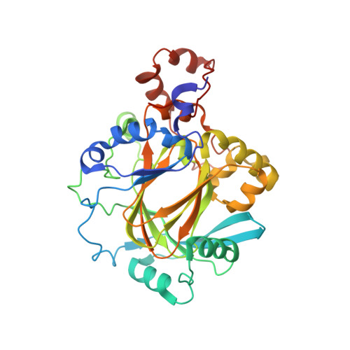
2Q8D

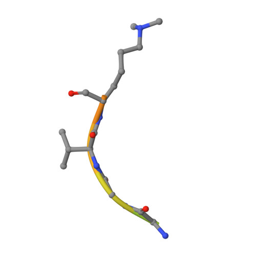
Crystal structure of JMJ2D2A in ternary complex with histone H3-K36me2 and succinate


Experimental Data Snapshot

  • Method: X-RAY DIFFRACTION
  • Resolution: 2.29 Å
  • R-Value Free: 0.251 
  • R-Value Work: 0.204 
  • R-Value Observed: 0.206 

Starting Model: experimental
View more details

wwPDB Validation   3D Report Full Report


This is version 1.4 of the entry. See complete history


Literature

Specificity and mechanism of JMJD2A, a trimethyllysine-specific histone demethylase.

Couture, J.F.Collazo, E.Ortiz-Tello, P.A.Brunzelle, J.S.Trievel, R.C.

(2007) Nat Struct Mol Biol 14: 689-695

  • DOI: https://doi.org/10.1038/nsmb1273
  • Primary Citation of Related Structures:  
    2Q8C, 2Q8D, 2Q8E

  • PubMed Abstract: 

    JMJD2A is a JmjC histone demethylase (HDM) that catalyzes the demethylation of di- and trimethylated Lys9 and Lys36 in histone H3 (H3K9me2/3 and H3K36me2/3). Here we present the crystal structures of the JMJD2A catalytic domain in complex with H3K9me3, H3K36me2 and H3K36me3 peptides. The structures reveal that histone substrates are recognized through a network of backbone hydrogen bonds and hydrophobic interactions that deposit the trimethyllysine into the active site. The trimethylated epsilon-ammonium cation is coordinated within a methylammonium-binding pocket through carbon-oxygen (CH...O) hydrogen bonds that position one of the zeta-methyl groups adjacent to the Fe(II) center for hydroxylation and demethylation. Mutations of the residues comprising this pocket abrogate demethylation by JMJD2A, with the exception of an S288A substitution, which augments activity, particularly toward H3K9me2. We propose that this residue modulates the methylation-state specificities of JMJD2 enzymes and other trimethyllysine-specific JmjC HDMs.


  • Organizational Affiliation

    Department of Biological Chemistry, University of Michigan, 1150 West Medical Center Drive, Ann Arbor, Michigan 48109-0606, USA.


Macromolecules
Find similar proteins by:  (by identity cutoff)  |  3D Structure
Entity ID: 1
MoleculeChains Sequence LengthOrganismDetailsImage
JmjC domain-containing histone demethylation protein 3A
A, B
352Homo sapiensMutation(s): 0 
Gene Names: JMJD2AJHDM3AJMJD2KIAA0677
EC: 1.14.11 (PDB Primary Data), 1.14.11.66 (UniProt), 1.14.11.69 (UniProt)
UniProt & NIH Common Fund Data Resources
Find proteins for O75164 (Homo sapiens)
Explore O75164 
Go to UniProtKB:  O75164
PHAROS:  O75164
GTEx:  ENSG00000066135 
Entity Groups  
Sequence Clusters30% Identity50% Identity70% Identity90% Identity95% Identity100% Identity
UniProt GroupO75164
Sequence Annotations
Expand
  • Reference Sequence

Find similar proteins by:  Sequence   |   3D Structure  

Entity ID: 2
MoleculeChains Sequence LengthOrganismDetailsImage
HISTONE 3 peptideC [auth F],
D [auth G]
16N/AMutation(s): 0 
Entity Groups  
Sequence Clusters30% Identity50% Identity70% Identity90% Identity95% Identity100% Identity
Sequence Annotations
Expand
  • Reference Sequence
Small Molecules
Modified Residues  1 Unique
IDChains TypeFormula2D DiagramParent
MLY
Query on MLY
C [auth F],
D [auth G]
L-PEPTIDE LINKINGC8 H18 N2 O2LYS
Experimental Data & Validation

Experimental Data

  • Method: X-RAY DIFFRACTION
  • Resolution: 2.29 Å
  • R-Value Free: 0.251 
  • R-Value Work: 0.204 
  • R-Value Observed: 0.206 
  • Space Group: P 21 21 2
Unit Cell:
Length ( Å )Angle ( ˚ )
a = 100.857α = 90
b = 148.642β = 90
c = 57.346γ = 90
Software Package:
Software NamePurpose
REFMACrefinement
Blu-Icedata collection
HKL-2000data reduction
HKL-2000data scaling
MOLREPphasing

Structure Validation

View Full Validation Report


Entry History 

Deposition Data

Revision History  (Full details and data files)

  • Version 1.0: 2007-07-03
    Type: Initial release
  • Version 1.1: 2008-05-01
    Changes: Version format compliance
  • Version 1.2: 2011-07-13
    Changes: Version format compliance
  • Version 1.3: 2018-01-24
    Changes: Structure summary
  • Version 1.4: 2023-08-30
    Changes: Data collection, Database references, Derived calculations, Refinement description