2QDG

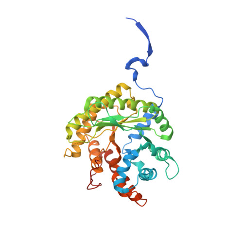
Fructose-1,6-bisphosphate Schiff base intermediate in FBP aldolase from Leishmania mexicana


Experimental Data Snapshot

  • Method: X-RAY DIFFRACTION
  • Resolution: 2.20 Å
  • R-Value Free: 0.238 
  • R-Value Work: 0.185 

Starting Model: experimental
View more details

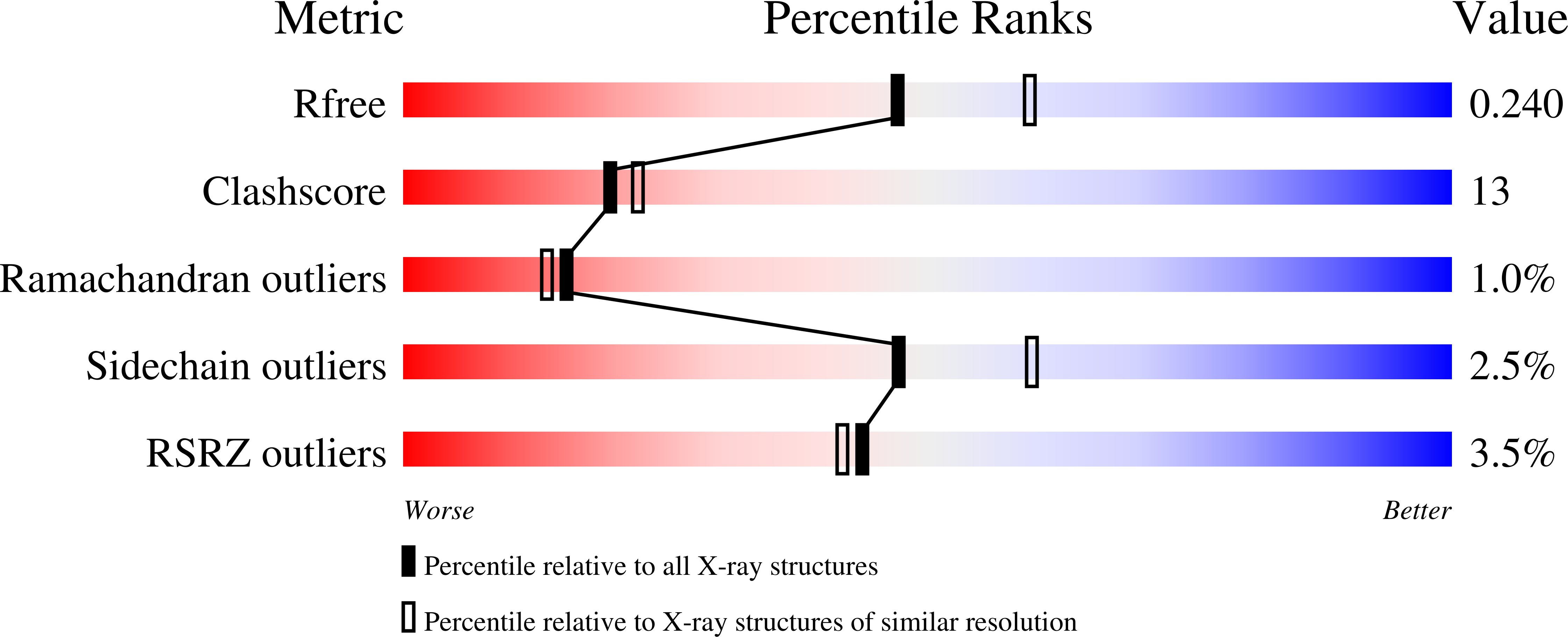
wwPDB Validation   3D Report Full Report


Ligand Structure Quality Assessment 


This is version 1.4 of the entry. See complete history


Literature

Carboxy-Terminus Recruitment Induced by Substrate Binding in Eukaryotic Fructose Bis-phosphate Aldolases

Lafrance-Vanasse, J.Sygusch, J.

(2007) Biochemistry 46: 9533-9540

  • DOI: https://doi.org/10.1021/bi700615r
  • Primary Citation of Related Structures:  
    2QAP, 2QDG, 2QDH

  • PubMed Abstract: 

    The crystal structures of Leishmania mexicana fructose-1,6-bis(phosphate) aldolase in complex with substrate and competitive inhibitor, mannitol-1,6-bis(phosphate), were solved to 2.2 A resolution. Crystallographic analysis revealed a Schiff base intermediate trapped in the native structure complexed with substrate while the inhibitor was trapped in a conformation mimicking the carbinolamine intermediate. Binding modes corroborated previous structures reported for rabbit muscle aldolase. Amino acid substitution of Gly-312 to Ala, adjacent to the P1-phosphate binding site and unique to trypanosomatids, did not perturb ligand binding in the active site. Ligand attachment ordered amino acid residues 359-367 of the C-terminal region (353-373) that was disordered beyond Asp-358 in the unbound structure, revealing a novel recruitment mechanism of this region by aldolases. C-Terminal peptide ordering is triggered by P1-phosphate binding that induces conformational changes whereby C-terminal Leu-364 contacts P1-phosphate binding residue Arg-313. C-Terminal region capture synergizes additional interactions with subunit surface residues, not perturbed by P1-phosphate binding, and stabilizes C-terminal attachment. Amino acid residues that participate in the capturing interaction are conserved among class I aldolases, indicating a general recruitment mechanism whereby C-terminal capture facilitates active site interactions in subsequent catalytic steps. Recruitment accelerates the enzymatic reaction by using binding energy to reduce configurational entropy during catalysis thereby localizing the conserved C-terminus tyrosine, which mediates proton transfer, proximal to the active site enamine.


  • Organizational Affiliation

    Département de Biochimie, Université de Montréal, Montréal, Québec H3C 3J7, Canada.


Macromolecules
Find similar proteins by:  (by identity cutoff)  |  3D Structure
Entity ID: 1
MoleculeChains Sequence LengthOrganismDetailsImage
Fructose-1,6-bisphosphate aldolase
A, B, C, D
391Leishmania mexicanaMutation(s): 0 
Gene Names: ald
EC: 4.1.2.13
UniProt
Find proteins for Q9U5N6 (Leishmania mexicana)
Explore Q9U5N6 
Go to UniProtKB:  Q9U5N6
Entity Groups  
Sequence Clusters30% Identity50% Identity70% Identity90% Identity95% Identity100% Identity
UniProt GroupQ9U5N6
Sequence Annotations
Expand
  • Reference Sequence
Small Molecules
Ligands 2 Unique
IDChains Name / Formula / InChI Key2D Diagram3D Interactions
2FP
Query on 2FP

Download Ideal Coordinates CCD File 
G [auth A],
J [auth B],
M [auth C],
P [auth D]
1,6-FRUCTOSE DIPHOSPHATE (LINEAR FORM)
C6 H14 O12 P2
XPYBSIWDXQFNMH-PYWDMBMJSA-N
PO4
Query on PO4

Download Ideal Coordinates CCD File 
E [auth A]
F [auth A]
H [auth B]
I [auth B]
K [auth C]
E [auth A],
F [auth A],
H [auth B],
I [auth B],
K [auth C],
L [auth C],
N [auth D],
O [auth D]
PHOSPHATE ION
O4 P
NBIIXXVUZAFLBC-UHFFFAOYSA-K
Experimental Data & Validation

Experimental Data

  • Method: X-RAY DIFFRACTION
  • Resolution: 2.20 Å
  • R-Value Free: 0.238 
  • R-Value Work: 0.185 
  • Space Group: P 21 21 21
Unit Cell:
Length ( Å )Angle ( ˚ )
a = 85.109α = 90
b = 117.09β = 90
c = 161.798γ = 90
Software Package:
Software NamePurpose
DENZOdata reduction
SCALEPACKdata scaling
CNSrefinement
PDB_EXTRACTdata extraction
CBASSdata collection
HKL-2000data reduction
HKL-2000data scaling
CNSphasing

Structure Validation

View Full Validation Report


Ligand Structure Quality Assessment 


Entry History 

Deposition Data

Revision History  (Full details and data files)

  • Version 1.0: 2007-08-21
    Type: Initial release
  • Version 1.1: 2011-07-13
    Changes: Derived calculations, Version format compliance
  • Version 1.2: 2017-10-18
    Changes: Refinement description
  • Version 1.3: 2023-08-30
    Changes: Data collection, Database references, Derived calculations, Refinement description
  • Version 1.4: 2024-11-20
    Changes: Structure summary