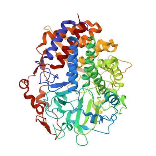
2QNO

Crystal Structure of the Mutant E55Q of the Cellulase CEL48F in Complex with a Thio-Oligosaccharide


Experimental Data Snapshot

  • Method: X-RAY DIFFRACTION
  • Resolution: 2.00 Å
  • R-Value Free: 0.182 
  • R-Value Work: 0.137 
  • R-Value Observed: 0.140 

Starting Model: experimental
View more details

wwPDB Validation   3D Report Full Report


This is version 2.2 of the entry. See complete history


Literature

Structures of mutants of cellulase Cel48F of Clostridium cellulolyticum in complex with long hemithiocellooligosaccharides give rise to a new view of the substrate pathway during processive action

Parsiegla, G.Reverbel, C.Tardif, C.Driguez, H.Haser, R.

(2008) J Mol Biol 375: 499-510

  • DOI: https://doi.org/10.1016/j.jmb.2007.10.039
  • Primary Citation of Related Structures:  
    1G9G, 1G9J, 2QNO

  • PubMed Abstract: 

    An efficient breakdown of lignocellulosic biomass is a prerequisite for the production of second-generation biofuels. Cellulases are key enzymes in this process. We crystallized complexes between hemithio-cello-deca and dodecaoses and the inactive mutants E44Q and E55Q of the endo-processive cellulase Cel48F, one of the most abundant cellulases in cellulosomes from Clostridium cellulolyticum, to elucidate its processive mechanism. In both complexes, the cellooligosaccharides occupy similar positions in the tunnel part of the active site but are more or less buried into the cleft, which hosts the active site. In the E44Q complex, it proceeds along the upper part of the cavity, while it occupies in the E55Q complex the same productive binding subsites in the lower part of the cavity that have previously been reported in Cel48F/cellooligosaccharide complexes. In both cases, the sugar moieties are stabilized by stacking interactions with aromatic side chains and H bonds. The upper pathway is gated by Tyr403, which blocks its access in the E55Q complex and offers a new stacking interaction in the E44Q complex. The new structural data give rise to the hypothesis of a two-step mechanism in which processive action and chain disruption occupy different subsites at the end of their trajectory. In the first part of the mechanism, the chain may smoothly slide up to the leaving group site along the upper pathway, while in the second part, the chain is cleaved in the already described productive binding position located in the lower pathway. The solved native structure of Cel48F without any bound sugar in the active site confirms the two side-chain orientations of the proton donor Glu55 as observed in the complex structures.


  • Organizational Affiliation

    Laboratoire de l'Architecture et Fonction des Macromolecules Biologiques, UMR 6098 CNRS and University of Aix-Marseille, Parc Scientifique et Technologique de Luminy, 13288 Marseille Cedex 09, France. [email protected]


Macromolecules
Find similar proteins by:  (by identity cutoff)  |  3D Structure
Entity ID: 1
MoleculeChains Sequence LengthOrganismDetailsImage
Endoglucanase F629Ruminiclostridium cellulolyticumMutation(s): 1 
Gene Names: celCCF
EC: 3.2.1.4
UniProt
Find proteins for P37698 (Ruminiclostridium cellulolyticum (strain ATCC 35319 / DSM 5812 / JCM 6584 / H10))
Explore P37698 
Go to UniProtKB:  P37698
Entity Groups  
Sequence Clusters30% Identity50% Identity70% Identity90% Identity95% Identity100% Identity
UniProt GroupP37698
Sequence Annotations
Expand
  • Reference Sequence
Oligosaccharides

Help

Entity ID: 2
MoleculeChains Length2D Diagram Glycosylation3D Interactions
4-thio-beta-D-glucopyranose-(1-4)-beta-D-glucopyranose-(1-4)-4-thio-beta-D-glucopyranose-(1-4)-beta-D-glucopyranose-(1-4)-4-thio-beta-D-glucopyranose-(1-4)-beta-D-glucopyranose-(1-4)-4-thio-beta-D-glucopyranose-(1-4)-beta-D-glucopyranose-(1-4)-4-thio-beta-D-glucopyranose
B
9N/AN/A
Glycosylation Resources
GlyTouCan:  G59113TZ
GlyCosmos:  G59113TZ
Small Molecules
Ligands 1 Unique
IDChains Name / Formula / InChI Key2D Diagram3D Interactions
CA
Query on CA

Download Ideal Coordinates CCD File 
C [auth A]CALCIUM ION
Ca
BHPQYMZQTOCNFJ-UHFFFAOYSA-N
Experimental Data & Validation

Experimental Data

  • Method: X-RAY DIFFRACTION
  • Resolution: 2.00 Å
  • R-Value Free: 0.182 
  • R-Value Work: 0.137 
  • R-Value Observed: 0.140 
  • Space Group: P 21 21 21
Unit Cell:
Length ( Å )Angle ( ˚ )
a = 61.24α = 90
b = 84.72β = 90
c = 121.74γ = 90
Software Package:
Software NamePurpose
SCALAdata scaling
REFMACrefinement
PDB_EXTRACTdata extraction
MAR345dtbdata collection
DENZOdata reduction
AMoREphasing

Structure Validation

View Full Validation Report


Entry History 

Deposition Data

Revision History  (Full details and data files)

  • Version 1.0: 2008-01-15
    Type: Initial release
  • Version 1.1: 2011-07-13
    Changes: Version format compliance
  • Version 2.0: 2020-07-29
    Type: Remediation
    Reason: Carbohydrate remediation
    Changes: Atomic model, Data collection, Derived calculations, Structure summary
  • Version 2.1: 2021-10-20
    Changes: Database references, Structure summary
  • Version 2.2: 2023-08-30
    Changes: Data collection, Refinement description