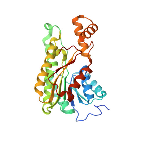
2RHC

Actinorhodin ketordeuctase, actKR, with NADP+ and Inhibitor Emodin


Experimental Data Snapshot

  • Method: X-RAY DIFFRACTION
  • Resolution: 2.10 Å
  • R-Value Free: 0.214 
  • R-Value Work: 0.186 

wwPDB Validation   3D Report Full Report


Ligand Structure Quality Assessment 


This is version 1.3 of the entry. See complete history


Literature

Inhibition kinetics and emodin cocrystal structure of a type II polyketide ketoreductase

Korman, T.P.Tan, Y.H.Wong, J.Luo, R.Tsai, S.-C.

(2008) Biochemistry 47: 1837-1847

  • DOI: https://doi.org/10.1021/bi7016427
  • Primary Citation of Related Structures:  
    2RH4, 2RHC, 2RHR

  • PubMed Abstract: 

    Type II polyketides are a class of natural products that include pharmaceutically important aromatic compounds such as the antibiotic tetracycline and antitumor compound doxorubicin. The type II polyketide synthase (PKS) is a complex consisting of 5-10 standalone domains homologous to fatty acid synthase (FAS). Polyketide ketoreductase (KR) provides regio- and stereochemical diversity during the reduction. How the type II polyketide KR specifically reduces only the C9 carbonyl group is not well understood. The cocrystal structures of actinorhodin polyketide ketoreductase (actKR) bound with NADPH or NADP+ and the inhibitor emodin were solved with the wild type and P94L mutant of actKR, revealing the first observation of a bent p-quinone in an enzyme active site. Molecular dynamics simulation help explain the origin of the bent geometry. Extensive screening for in vitro substrates shows that unlike FAS KR, the actKR prefers bicyclic substrates. Inhibition kinetics indicate that actKR follows an ordered Bi Bi mechanism. Together with docking simulations that identified a potential phosphopantetheine binding groove, the structural and functional studies reveal that the C9 specificity is a result of active site geometry and substrate ring constraints. The results lay the foundation for the design of novel aromatic polyketide natural products with different reduction patterns.


  • Organizational Affiliation

    Department of Molecular Biology and Biochemistry, University of California, Irvine, California 92697, USA.


Macromolecules
Find similar proteins by:  (by identity cutoff)  |  3D Structure
Entity ID: 1
MoleculeChains Sequence LengthOrganismDetailsImage
Actinorhodin Polyketide KetoreductaseA [auth B],
B [auth A]
277Streptomyces coelicolorMutation(s): 0 
Gene Names: actIII
EC: 1.3.1
UniProt
Find proteins for P16544 (Streptomyces coelicolor (strain ATCC BAA-471 / A3(2) / M145))
Explore P16544 
Go to UniProtKB:  P16544
Entity Groups  
Sequence Clusters30% Identity50% Identity70% Identity90% Identity95% Identity100% Identity
UniProt GroupP16544
Sequence Annotations
Expand
  • Reference Sequence
Experimental Data & Validation

Experimental Data

  • Method: X-RAY DIFFRACTION
  • Resolution: 2.10 Å
  • R-Value Free: 0.214 
  • R-Value Work: 0.186 
  • Space Group: P 32 2 1
Unit Cell:
Length ( Å )Angle ( ˚ )
a = 103.875α = 90
b = 103.875β = 90
c = 123.283γ = 120
Software Package:
Software NamePurpose
DENZOdata reduction
SCALEPACKdata scaling
CNSrefinement
PDB_EXTRACTdata extraction
ADSCdata collection
HKL-2000data reduction
HKL-2000data scaling
CNSphasing

Structure Validation

View Full Validation Report


Ligand Structure Quality Assessment 


Entry History 

Deposition Data

Revision History  (Full details and data files)

  • Version 1.0: 2008-08-19
    Type: Initial release
  • Version 1.1: 2011-07-13
    Changes: Version format compliance
  • Version 1.2: 2017-10-25
    Changes: Refinement description
  • Version 1.3: 2024-02-21
    Changes: Data collection, Database references, Derived calculations