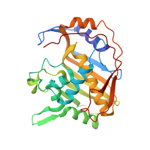
Structure, multiple site binding, and segmental accommodation in thymidylate synthase on binding dUMP and an anti-folate.
Montfort, W.R., Perry, K.M., Fauman, E.B., Finer-Moore, J.S., Maley, G.F., Hardy, L., Maley, F., Stroud, R.M.(1990) Biochemistry 29: 6964-6977
- PubMed: 2223754
- DOI: https://doi.org/10.1021/bi00482a004
- Primary Citation of Related Structures:
2TSC - PubMed Abstract:
The structure of Escherichia coli thymidylate synthase (TS) complexed with the substrate dUMP and an analogue of the cofactor methylenetetrahydrofolate was solved by multiple isomorphous replacement and refined at 1.97-A resolution to a residual of 18% for all data (16% for data greater than 2 sigma) for a highly constrained structure. All residues in the structure are clearly resolved and give a very high confidence in total correctness of the structure. The ternary complex directly suggests how methylation of dUMP takes place. C-6 of dUMP is covalently bound to gamma S of Cys-198(146) during catalysis, and the reactants are surrounded by specific hydrogen bonds and hydrophobic interactions from conserved residues. Comparison with the independently solved structure of unliganded TS reveals a large conformation change in the enzyme, which closes down to sequester the reactants and several highly ordered water molecules within a cavernous active center, away from bulk solvent. A second binding site for the quinazoline ring of the cofactor analogue was discovered by withholding addition of reducing agent during crystal storage. The chemical change in the protein is slight, and from difference density maps modification of sulfhydryls is not directly responsible for blockade of the primary site. The site, only partially overlapping with the primary site, is also surrounded by conserved residues and thus may play a functional role. The ligand-induced conformational change is not a domain shift but involves the segmental accommodation of several helices, beta-strands, and loops that move as units against the beta-sheet interface between monomers.
Organizational Affiliation:
Department of Biochemistry and Biophysics, University of California, San Francisco 94143-0448.


