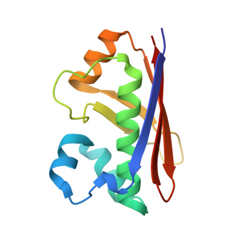
2WAT

Structure of the fungal type I FAS PPT domain in complex with CoA


Experimental Data Snapshot

  • Method: X-RAY DIFFRACTION
  • Resolution: 2.20 Å
  • R-Value Free: 0.252 
  • R-Value Work: 0.217 
  • R-Value Observed: 0.219 

wwPDB Validation   3D Report Full Report


Ligand Structure Quality Assessment 


This is version 1.3 of the entry. See complete history


Literature

Multimeric Options for the Auto-Activation of the Saccharomyces Cerevisiae Fas Type I Megasynthase.

Johansson, P.Mulinacci, B.Koestler, C.Vollrath, R.Oesterhelt, D.Grininger, M.

(2009) Structure 17: 1063

  • DOI: https://doi.org/10.1016/j.str.2009.06.014
  • Primary Citation of Related Structures:  
    2WAS, 2WAT, 3HMJ

  • PubMed Abstract: 

    The fungal type I fatty acid synthase (FAS) is a 2.6 MDa multienzyme complex, catalyzing all necessary steps for the synthesis of long acyl chains. To be catalytically competent, the FAS must be activated by a posttranslational modification of the central acyl carrier domain (ACP) by an intrinsic phosphopantetheine transferase (PPT). However, recent X-ray structures of the fungal FAS revealed a barrel-shaped architecture, with PPT located at the outside of the barrel wall, spatially separated from the ACP caged in the inner volume. This separation indicated that the activation has to proceed before the assembly to the mature complex, in a conformation where the ACP and PPT domains can meet. To gain insight into the auto-activation reaction and also into the fungal FAS assembly pathway, we structurally and functionally characterized the Saccharomyces cerevisiae FAS type I PPT as part of the multienzyme protein and as an isolated domain.


  • Organizational Affiliation

    Department of Membrane Biochemistry, Max-Planck-Institute of Biochemistry, Martinsried, Germany. [email protected]


Macromolecules
Find similar proteins by:  (by identity cutoff)  |  3D Structure
Entity ID: 1
MoleculeChains Sequence LengthOrganismDetailsImage
3-OXOACYL-[ACYL-CARRIER-PROTEIN] SYNTHASE
A, B, C, D, E
A, B, C, D, E, F
122Saccharomyces cerevisiaeMutation(s): 0 
EC: 2.7.8.7 (PDB Primary Data), 2.3.1.41 (PDB Primary Data), 2.3.1.86 (UniProt), 1.1.1.100 (UniProt)
UniProt
Find proteins for P19097 (Saccharomyces cerevisiae (strain ATCC 204508 / S288c))
Explore P19097 
Go to UniProtKB:  P19097
Entity Groups  
Sequence Clusters30% Identity50% Identity70% Identity90% Identity95% Identity100% Identity
UniProt GroupP19097
Sequence Annotations
Expand
  • Reference Sequence
Small Molecules
Ligands 3 Unique
IDChains Name / Formula / InChI Key2D Diagram3D Interactions
COA
Query on COA

Download Ideal Coordinates CCD File 
H [auth B],
I [auth B],
M [auth C],
Q [auth E],
R [auth E]
COENZYME A
C21 H36 N7 O16 P3 S
RGJOEKWQDUBAIZ-IBOSZNHHSA-N
CL
Query on CL

Download Ideal Coordinates CCD File 
G [auth A],
L [auth B],
P [auth D],
T [auth E]
CHLORIDE ION
Cl
VEXZGXHMUGYJMC-UHFFFAOYSA-M
MG
Query on MG

Download Ideal Coordinates CCD File 
J [auth B],
K [auth B],
N [auth C],
O [auth D],
S [auth E]
MAGNESIUM ION
Mg
JLVVSXFLKOJNIY-UHFFFAOYSA-N
Experimental Data & Validation

Experimental Data

  • Method: X-RAY DIFFRACTION
  • Resolution: 2.20 Å
  • R-Value Free: 0.252 
  • R-Value Work: 0.217 
  • R-Value Observed: 0.219 
  • Space Group: C 1 2 1
Unit Cell:
Length ( Å )Angle ( ˚ )
a = 145.587α = 90
b = 73.764β = 130.8
c = 105.689γ = 90
Software Package:
Software NamePurpose
PHENIXrefinement
PHASERphasing

Structure Validation

View Full Validation Report


Ligand Structure Quality Assessment 


Entry History 

Deposition Data

Revision History  (Full details and data files)

  • Version 1.0: 2009-08-25
    Type: Initial release
  • Version 1.1: 2011-05-08
    Changes: Version format compliance
  • Version 1.2: 2011-07-13
    Changes: Version format compliance
  • Version 1.3: 2024-05-08
    Changes: Data collection, Database references, Derived calculations, Other