Structural Basis for the Erythro-Stereospecificity of the L-Arginine Oxygenase Vioc in Viomycin Biosynthesis.
Helmetag, V., Samel, S.A., Thomas, M.G., Marahiel, M.A., Essen, L.-O.(2009) FEBS J 276: 3669
- PubMed: 19490124
- DOI: https://doi.org/10.1111/j.1742-4658.2009.07085.x
- Primary Citation of Related Structures:
2WBO, 2WBP, 2WBQ - PubMed Abstract:
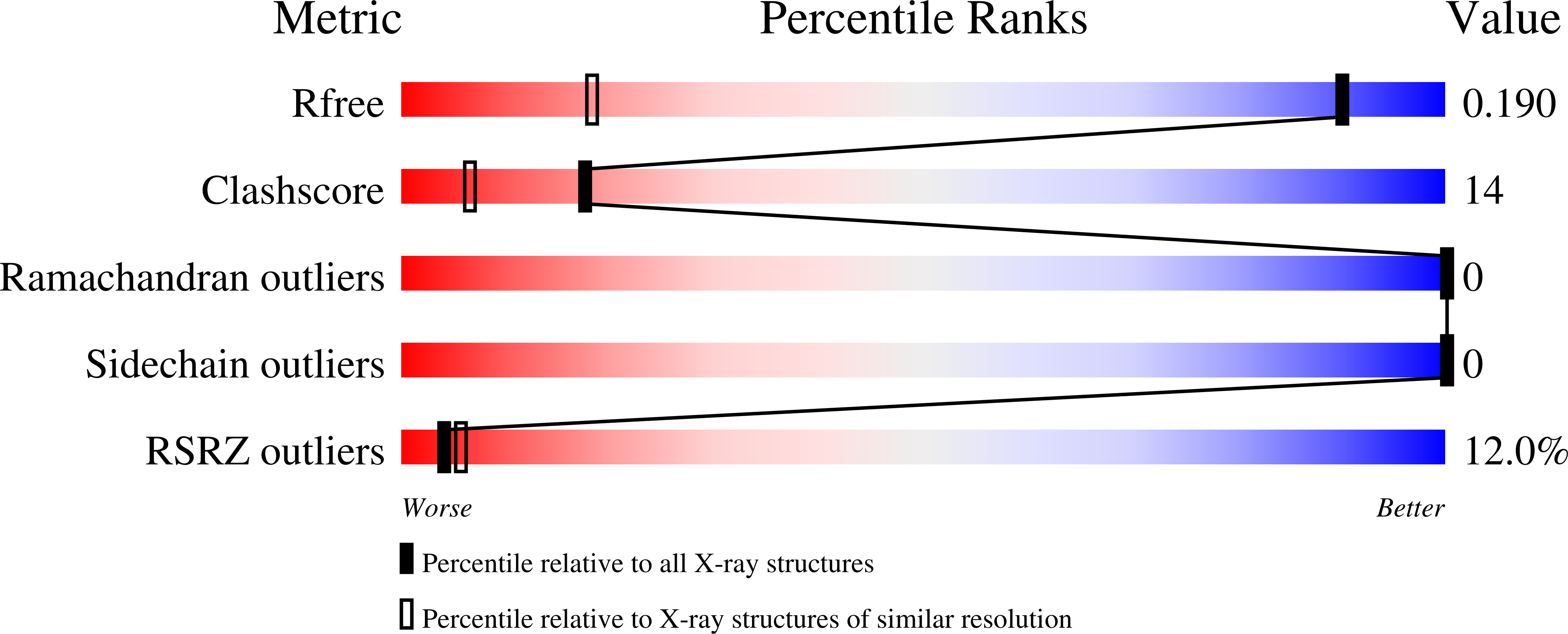
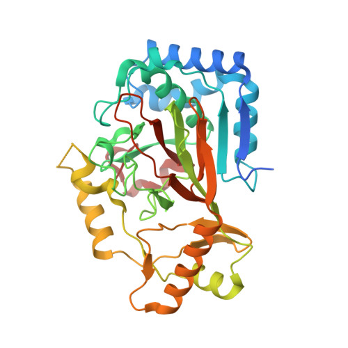
The nonheme iron oxygenase VioC from Streptomyces vinaceus catalyzes Fe(II)-dependent and alpha-ketoglutarate-dependent Cbeta-hydroxylation of L-arginine during the biosynthesis of the tuberactinomycin antibiotic viomycin. Crystal structures of VioC were determined in complexes with the cofactor Fe(II), the substrate L-arginine, the product (2S,3S)-hydroxyarginine and the coproduct succinate at 1.1-1.3 A resolution. The overall structure reveals a beta-helix core fold with two additional helical subdomains that are common to nonheme iron oxygenases of the clavaminic acid synthase-like superfamily. In contrast to other clavaminic acid synthase-like oxygenases, which catalyze the formation of threo diastereomers, VioC produces the erythro diastereomer of Cbeta-hydroxylated L-arginine. This unexpected stereospecificity is caused by conformational control of the bound substrate, which enforces a gauche(-) conformer for chi(1) instead of the trans conformers observed for the asparagine oxygenase AsnO and other members of the clavaminic acid synthase-like superfamily. Additionally, the substrate specificity of VioC was investigated. The side chain of the L-arginine substrate projects outwards from the active site by undergoing interactions mainly with the C-terminal helical subdomain. Accordingly, VioC exerts broadened substrate specificity by accepting the analogs L-homoarginine and L-canavanine for Cbeta-hydroxylation.
Organizational Affiliation:
Department of Chemistry, Philipps-University Marburg, Germany.


